Thomas A Roberts1, Laurence H Jackson1, Joshua FP van Amerom2, Alena Uus1, Anthony N Price1, Johannes K Steinweg1, David FA Lloyd1,3, Milou PM van Poppel1, Kuberan Pushparajah3, Mary A Rutherford1, Reza Razavi1,3, Maria Deprez1, and Joseph V Hajnal1
1Biomedical Engineering Department, School of Biomedical Engineering and Imaging Sciences, King's College London, London, United Kingdom, 2Division of Pediatric Cardiology, The Hospital for Sick Children, Toronto, ON, Canada, 3Department of Congenital Heart Disease, Evelina Children's Hospital, London, United Kingdom
1Biomedical Engineering Department, School of Biomedical Engineering and Imaging Sciences, King's College London, London, United Kingdom, 2Division of Pediatric Cardiology, The Hospital for Sick Children, Toronto, ON, Canada, 3Department of Congenital Heart Disease, Evelina Children's Hospital, London, United Kingdom
We combine k-t SENSE with a SWEEP excitation to perform faster fetal whole-heart anatomical and blood flow 4D cine MRI. In combination with self-calibrated k-t methods, this framework paves the way for development of comprehensive clinical 4D fetal whole heart MRI in under five minutes.
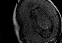
Figure 3: Movie showing every fourth image frame from a k-t SWEEP acquisition acquired in a fetus aged 30+2 weeks gestational age.The k-t SWEEP acquisition smoothly sweeps across the field-of-view (compare against the k-t M2D movie in Fig 2). In the k-t SWEEP data a stable signal is reached after an initial transient period, which manifests as noisy, fluctuating image frames at the beginning of the movie. Note: image quality not fully representative of scan quality due to the need for extreme gif compression.
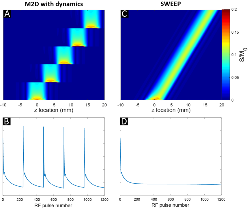
Figure 1: Simulations of M2D and SWEEP [6] acquisition schemes. In both, the same number of images are acquired and coverage in the z-direction is equivalent. For the M2D acquisition (A), multiple dynamics are acquired for each slice z-location. (B) For each slice, 100s of RF pulses are required before the steady-state signal is reached. With SWEEP, (C) the excitation frequency of the RF pulse is slowly linearly increased resulting in a gradually shifting slice profile across the FOV. (D) After the initial transient period, a stable signal results throughout the rest of the acquisition.