Zhixing Wang1, Steven Allen1, Xue Feng1, John P. Mugler2, and Craig H. Meyer1
1Biomedical Engineering, University of Virginia, Charlottesville, VA, United States, 2Radiology & Medical Imaging, University of Virginia, Charlottesville, VA, United States
1Biomedical Engineering, University of Virginia, Charlottesville, VA, United States, 2Radiology & Medical Imaging, University of Virginia, Charlottesville, VA, United States
This study presents a new approach to 2D TSE imaging using annular spiral rings with a retraced in/out trajectory, dubbed “SPRING-RIO TSE”, for fast, high-quality, T2-weighted brain imaging with higher scan efficiency and reduced SAR, when compared with Cartesian TSE imaging.
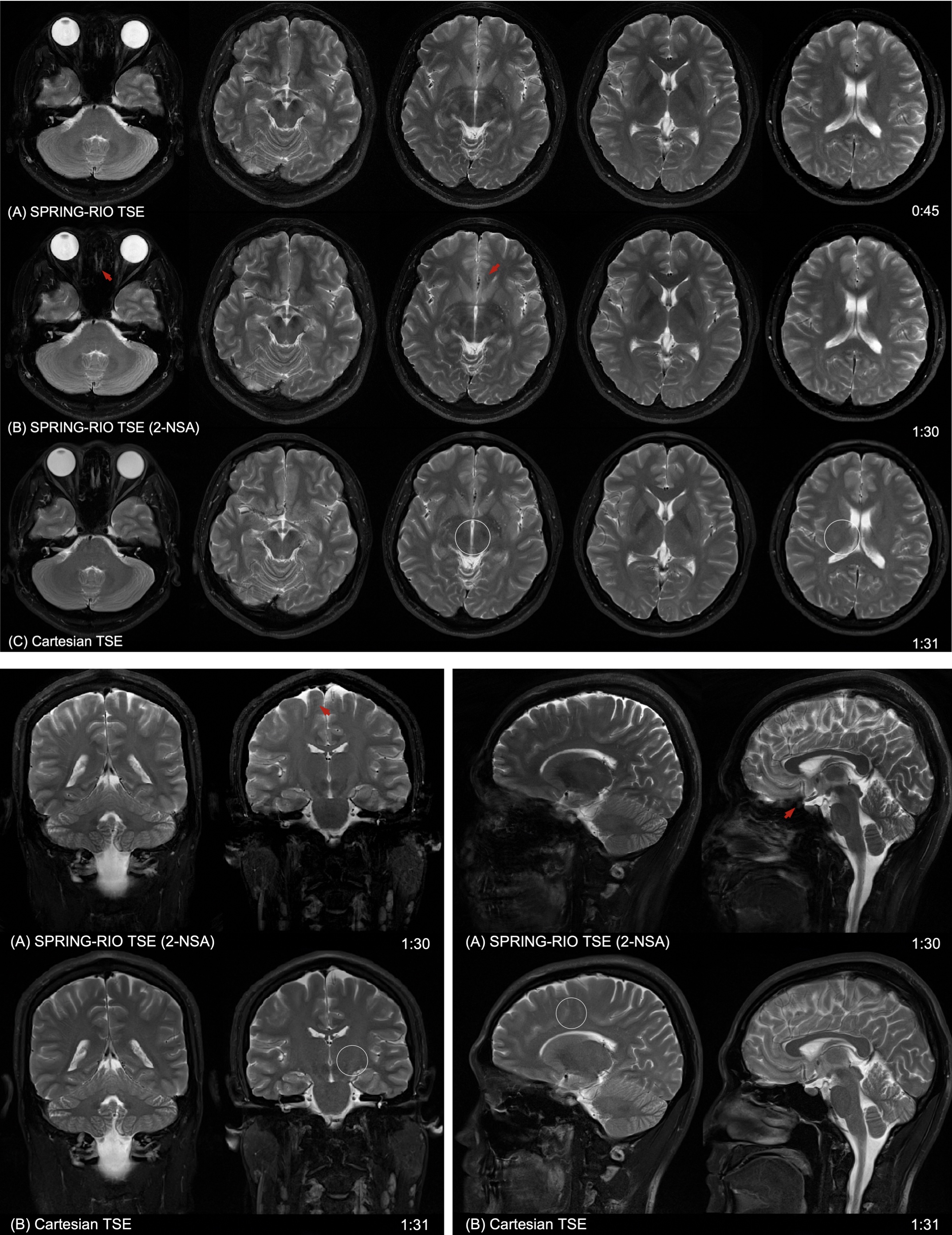
Figure 4. Comparison of in-vivo images acquired using the proposed SPRING-RIO TSE method and standard Cartesian TSE. From top to bottom are trajectory- and off-resonance-corrected images from SPRING-RIO TSE with one signal average and with two signal averages, and images from standard Cartesian TSE. The red arrows point to structures where residual signal loss or artifacts exist, likely due to strong susceptibility, concomitant gradients or flow effects. The white circles indicate the spots where the image contrast is visually better in SPRING-RIO TSE than that in Cartesian TSE.
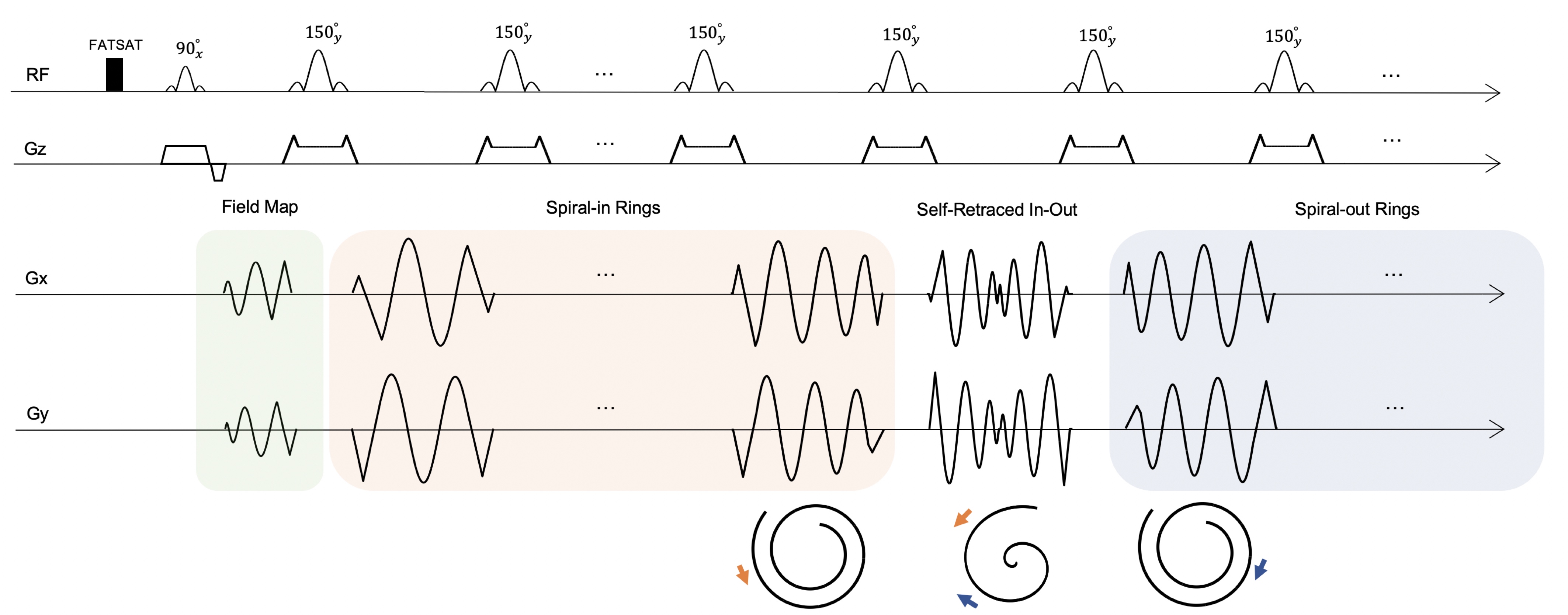
Figure 1. Pulse sequence diagram showing the sampling strategy, which includes fat saturation, field-map acquisition, and data acquisition using annular spiral rings. The center of k space is sampled by a self-retraced spiral in-out arm. The spiral-in rings in the orange box are designed to cover the outer k space, while the spiral-out rings in the blue box retrace the corresponding spiral-in rings. The refocusing RF pulse angles are set to 150° for reducing SAR.