Myung-Ho In1, Norbert G Campeau1, John III Huston1, Zijing Dong2,3, Kawin Setsompop4,5, Daehun Kang1, Uten Yarach6, Yunhong Shu1, Joshua D Trzasko1, and Matt A Bernstein1
1Department of Radiology, Mayo Clinic, Rochester, MN, United States, 2A. A. Martinos Center for Biomedical Imaging, Department of Radiology, Massachusetts General Hospital, Charlestown, MA, United States, 3Department of Electrical Engineering and Computer Science, MIT, Cambridge, MA, United States, 4Department of Radiology, Stanford University, Stanford, CA, United States, 5Stanford University, Stanford, CA, United States, 6Department of Radiologic Technology, Faculty of Associated Medical Sciences, Chiang Mai University, Chiang Mai, Thailand
1Department of Radiology, Mayo Clinic, Rochester, MN, United States, 2A. A. Martinos Center for Biomedical Imaging, Department of Radiology, Massachusetts General Hospital, Charlestown, MA, United States, 3Department of Electrical Engineering and Computer Science, MIT, Cambridge, MA, United States, 4Department of Radiology, Stanford University, Stanford, CA, United States, 5Stanford University, Stanford, CA, United States, 6Department of Radiologic Technology, Faculty of Associated Medical Sciences, Chiang Mai University, Chiang Mai, Thailand
DIADEM, a variant of
multi-shot EPI, was optimized and evaluated as a high-speed alternative to T2-FSE
in 24 subjects. Overall T2-DIADEM performed well relative to T2-FSE in terms of
resolution and contrast, but suffered more from flow related artifacts, signal
dropout and Gibbs ringing.
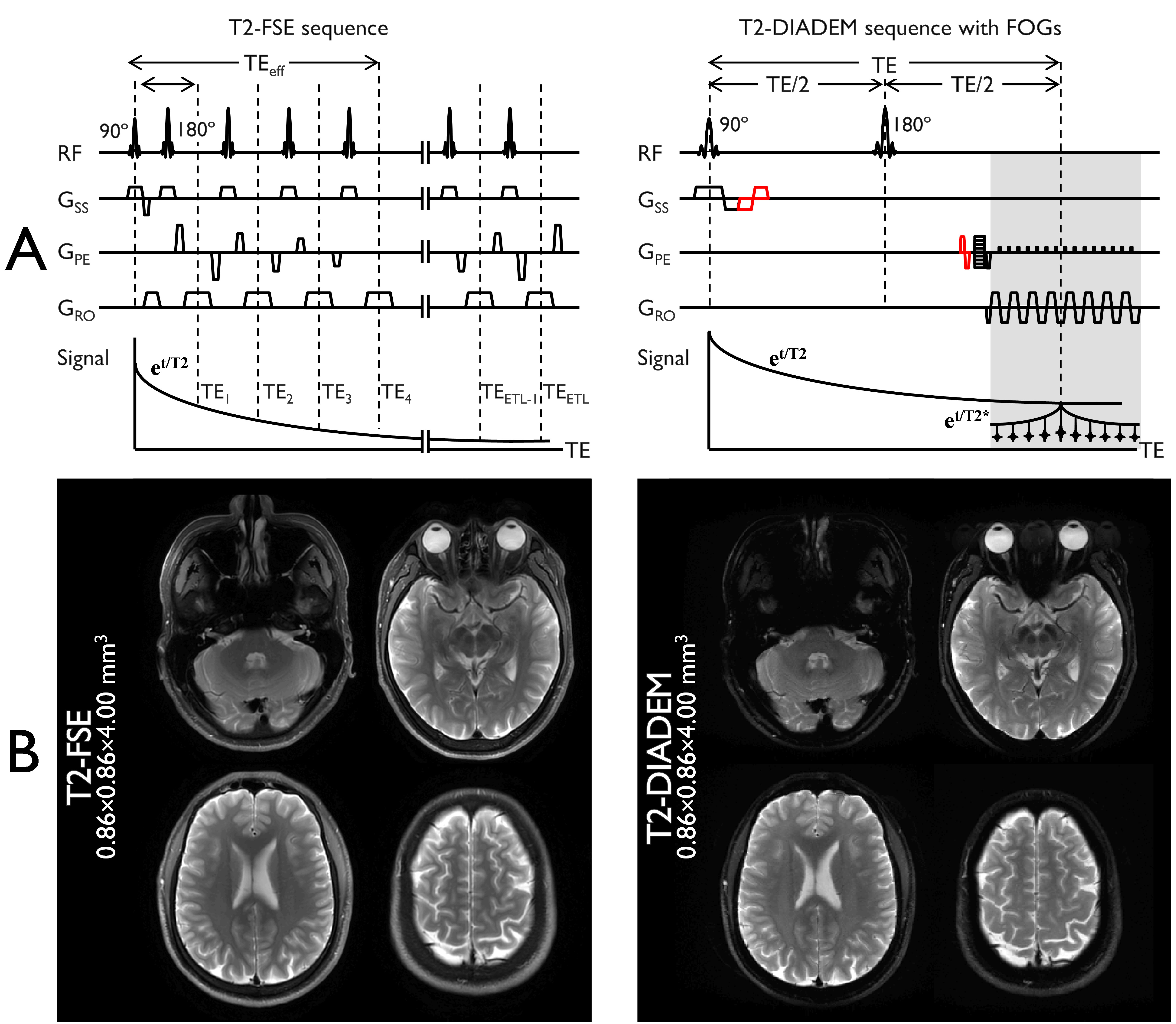
Figure 2 Imaging sequences of
T2-FSE and T2-DIADEM (A) and corresponding images (B). The flow compensation
gradients on the DIADEM sequence are shown in red-colors in (A). For
demonstration purpose, four slices among 38 are shown in (B).
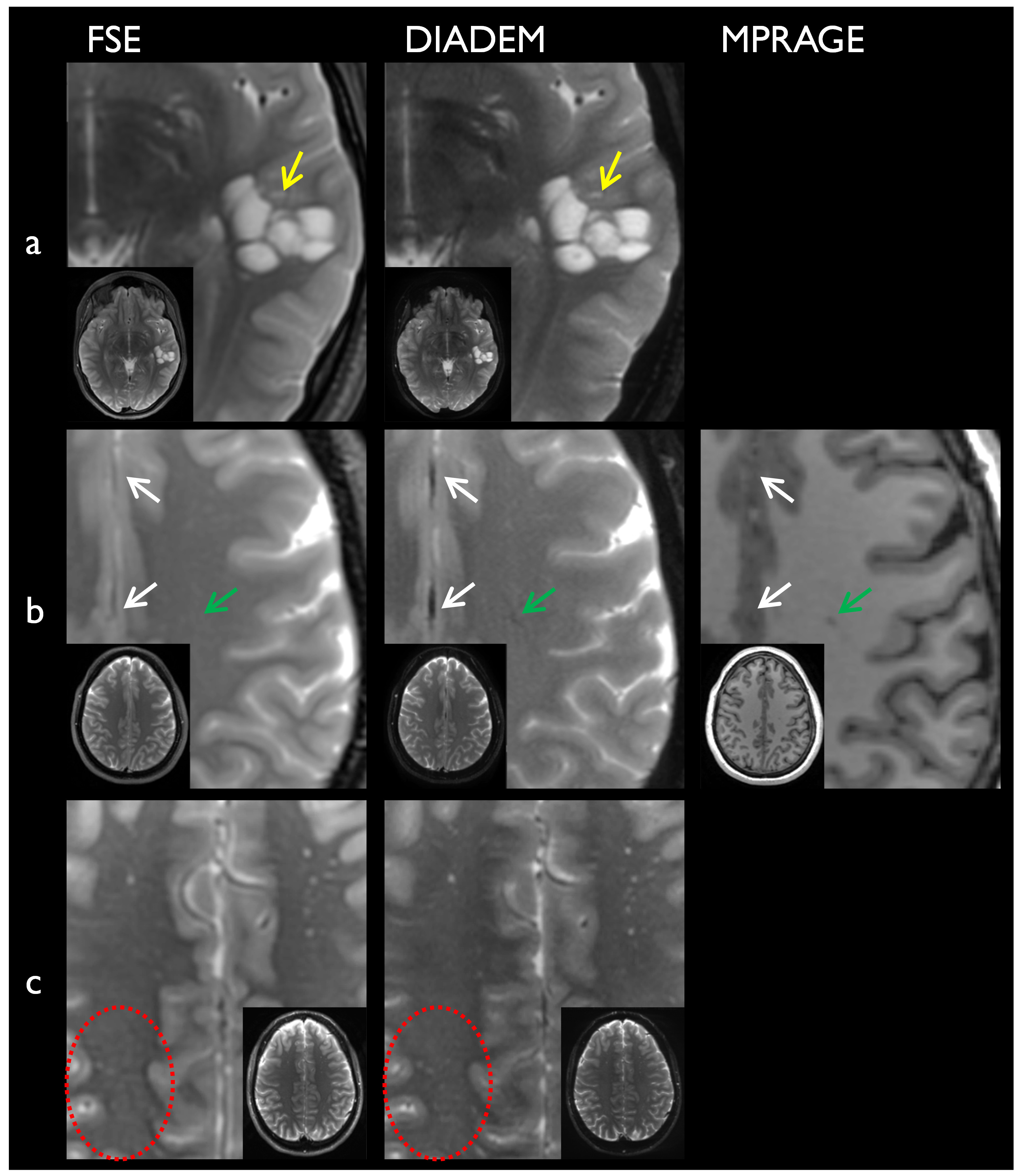
Figure 3. Advantages of
T2-DIADEM over T2-FSE: T2-DIADEM provides better depiction of internal details
of left temporal lobe multi-cystic lesion (a), falx calcifications (white
arrows in b), small T2 hyperintensities (c). T2-DIADEM identifies small
developmental venous anomaly, as demonstrated in MPRAGE (green arrows in b)