Edwin Versteeg1, Sarah M. Jacobs1, Ícaro A.F. Oliveira2, Dennis W.J. Klomp1, and Jeroen C.W. Siero1,2
1Radiology, University Medical Center Utrecht, Utrecht, Netherlands, 2Spinoza centre for neuroimaging Amsterdam, Amsterdam, Netherlands
1Radiology, University Medical Center Utrecht, Utrecht, Netherlands, 2Spinoza centre for neuroimaging Amsterdam, Amsterdam, Netherlands
In this work, we implemented a silent readout module that applies a silent gradient axis (driven @20kHz) to a 3D MPRAGE sequence. The resulting sequence featured a 26 dB reduction in peak sound level and comparable image contrast and scan time compared to a conventional 3D MPRAGE-scan.
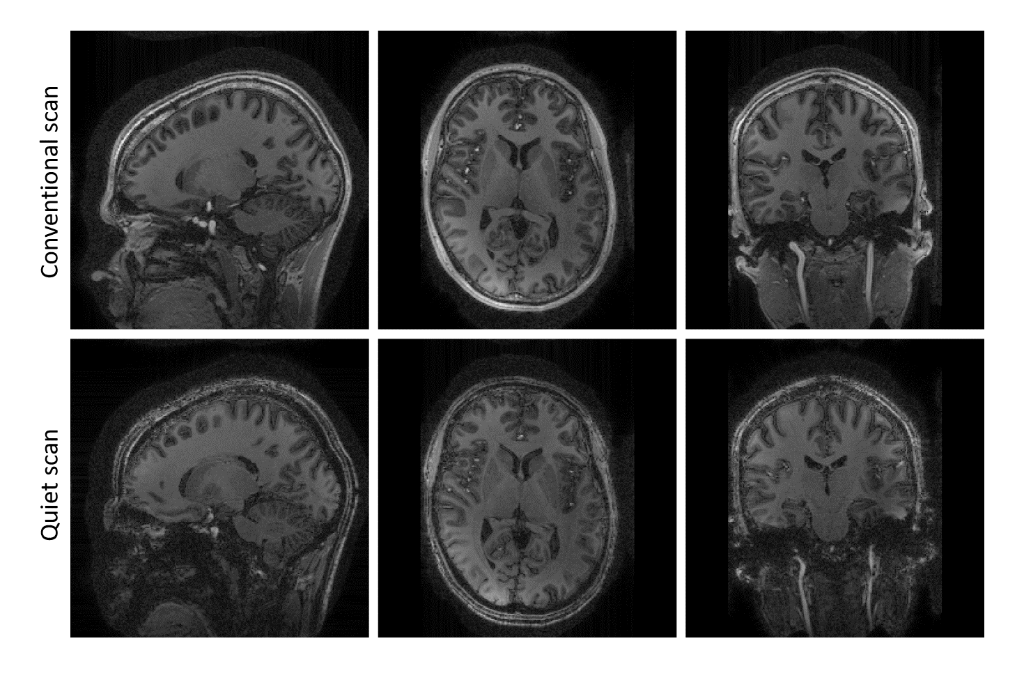
Figure
3: Representative slices of the whole brain MPRAGE
scans. Note that the same windowing is used for all images, which highlight the
similar contrast between the conventional (top row) and quiet scan (bottom
row). A slight misalignment between
anatomy might be visible due to subject movement.
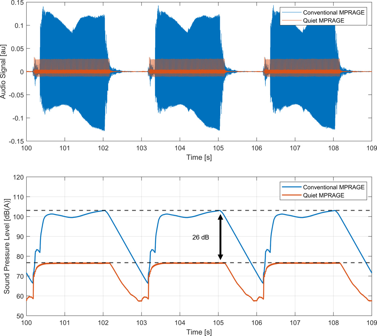
Figure
4: Part of the sound measurement used to estimate
the sound levels. Here, three shots of the MPRAGE-scans are displayed. Top: the
audio signal from the microphone after A-weighting. Bottom: the calculated
sound pressure levels (dB(A) and fast
time weighting) for each shot.