Takayuki Sakai1,2, Masami Yoneyama3, Atsuya Watanabe4,5, Daichi Murayama1, Shigehiro Ochi1, Shuo Zhang6, and Tosiaki Miyati7
1Radiology, Eastern Chiba Medical Center, Tonage, Japan, 2Division of Health Sciences, Graduate School of Medical Sciences, Kanazawa University, Kanazawa, Japan, 3Philips Japan, Tokyo, Japan, 4General Medical Services, Chiba University Graduate School of Medicine, Chiba, Japan, 5Orthopaedic Surgery, Eastern Chiba Medical Center, Chiba, Japan, 6Philips Healthcare, Hamburg, Germany, 7Faculty of Health Sciences, Institute of Medical, Pharmaceutical and Health Sciences, Kanazawa University, Kanazawa, Japan
1Radiology, Eastern Chiba Medical Center, Tonage, Japan, 2Division of Health Sciences, Graduate School of Medical Sciences, Kanazawa University, Kanazawa, Japan, 3Philips Japan, Tokyo, Japan, 4General Medical Services, Chiba University Graduate School of Medicine, Chiba, Japan, 5Orthopaedic Surgery, Eastern Chiba Medical Center, Chiba, Japan, 6Philips Healthcare, Hamburg, Germany, 7Faculty of Health Sciences, Institute of Medical, Pharmaceutical and Health Sciences, Kanazawa University, Kanazawa, Japan
MIXTURE successful offers multi-contrast and quantitative images within one single scan, which could provide information for potential anatomical and pathological assessment simultaneously for knee imaging.
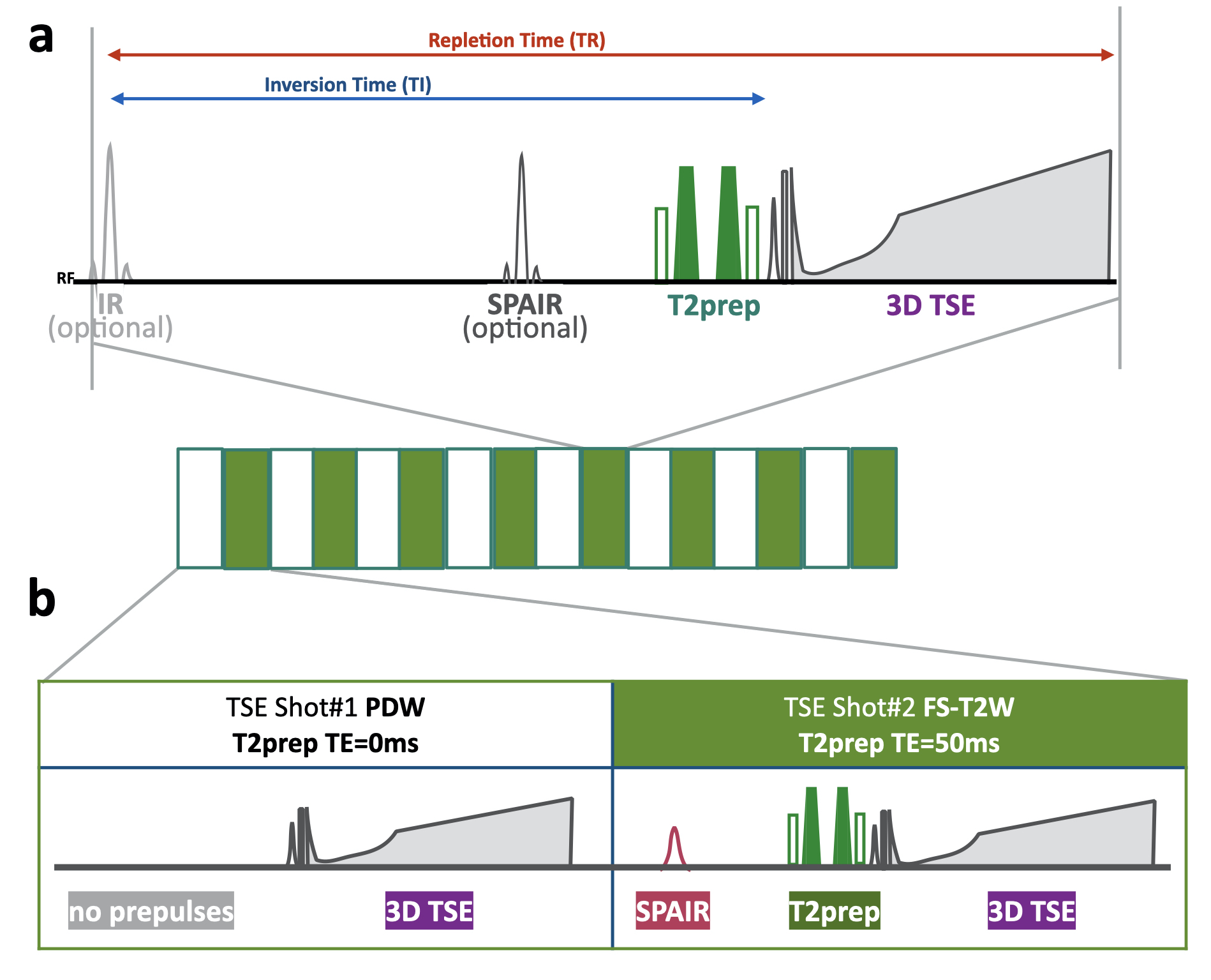
Fig.1 Scheme of the MIXTURE (Multi-Interleaved X-prepared tse with inTUitive RElaxometry).
(a) T2-mapping was performed using T2-prepared 3D segmented turbo spin- echo (TSE) with variable refocusing pulse trains.(b) Two images with different TE (TE = 0 and 50ms) were acquired with interleaved acquisition. To obtain the compatible contrasts with routine TSE images, TSE shot#1 did not apply any pre-pulses (as “PDW) and shot#2 applied both SPAIR and T2pre (as “fat-suppressed T2W”).
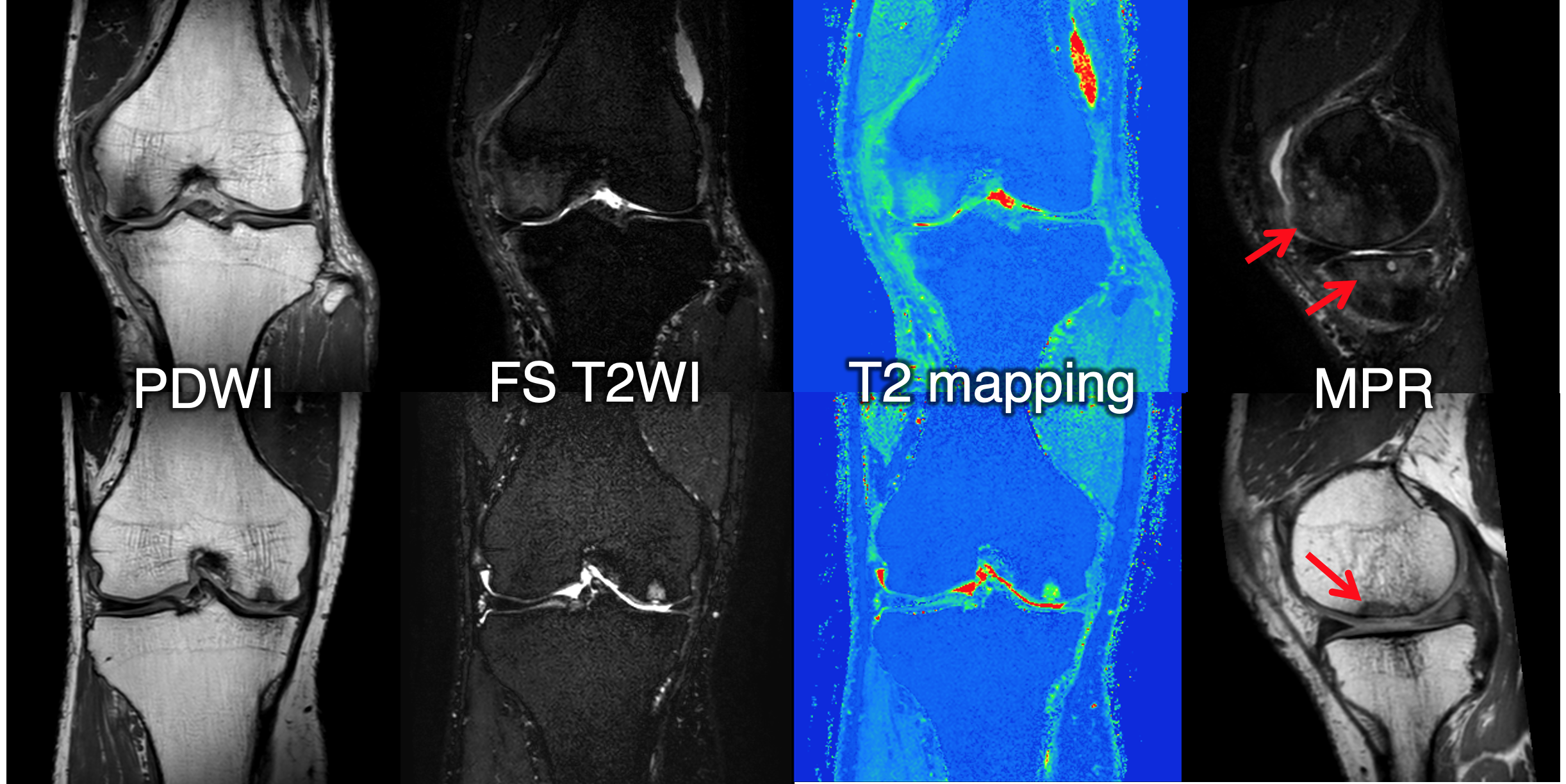
Fig.4 Simultaneous multi-contrast and quantitative images of MIXTURE from a patient with knee joint pain.
Upper: 57y male, knee osteoarthritis, osteonecrosis, bone bruise, intraosseous cyst.Lower: 65y male, idiopathic osteonecrosis.