Guangtao Fan1, Yudan Li1, Fenglin Xue2, Yilong Huang1, Yanlin Li3, Guoliang Wang3, Tianfu Qi1, Lisha Nie4, and Bo He1
1Department of Imaging, the First Affiliated Hospital of Kunming Medical University, Kunming, China, 2Department of Pathology, the First Affiliated Hospital of Kunming Medical University, Kunming, China, 3Department of Sports Medicine, the First Affiliated Hospital of Kunming Medical University, Kunming, China, 4GE Healthcare, Kunming, China
1Department of Imaging, the First Affiliated Hospital of Kunming Medical University, Kunming, China, 2Department of Pathology, the First Affiliated Hospital of Kunming Medical University, Kunming, China, 3Department of Sports Medicine, the First Affiliated Hospital of Kunming Medical University, Kunming, China, 4GE Healthcare, Kunming, China
The study aims to explore the clinical application value of MRI to
quantitatively assess the ACL-MD. It was concluded that MRI T1, T2, T2* values can
quantitatively evaluate knee joint ACL-MD, and T2* value has the highest
diagnostic efficiency.
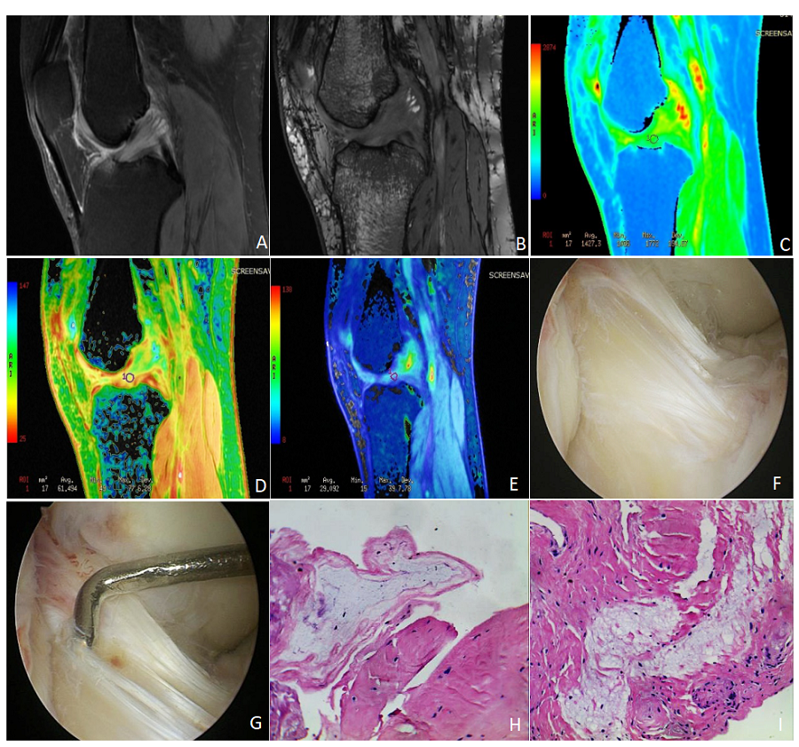
Figure 1: Images of an ACL-MD patient who is a 61-year-old female with the OSag fs (Oblique Sagittal fat suppression Proton Density weighted image), sagittal 3D-FIESTA, T1mapping, T2mapping, T2*mapping, arthroscopy and pathology.
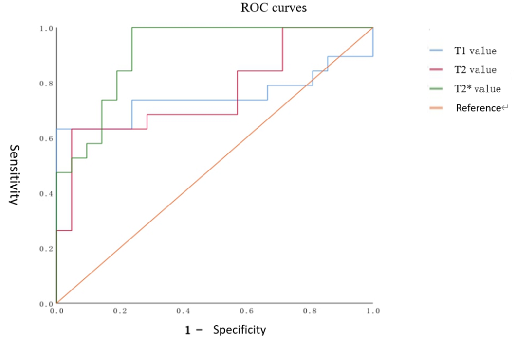
Figure 2: The curves of T1, T2, and T2* values in the ACL-MD group.