Jikai Shen1, Qi Zhao1, Yi Qi1, Gary Cofer1, G. Allan Johnson1, and Nian Wang2
1Duke University, Durham, NC, United States, 2Radiology and Imaging Sciences, Indiana University, Indianapolis, IN, United States
1Duke University, Durham, NC, United States, 2Radiology and Imaging Sciences, Indiana University, Indianapolis, IN, United States
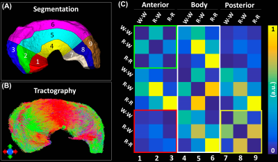
Strong zonal-dependent diffusion properties were demonstrated by DTI metrics (FA, MD, AD, and RD). Combining tractography and automatic segmentation method, we were able to observe the structural connections among different areas of the meniscus.

Figure 5. The structural connection heatmap of meniscus (c) obtained by the automatic parcellation (a) and tractography (b).

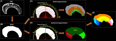
Figure 1. The automatic segmentation process used in this study, from the acquired DWI (a) to the 9 different areas of meniscus (e). Both Radial Segmentation (c) and Rotational Segmentation (d) were derived from the binary mask (b). These two methods were further combined to divide the whole meniscus to 9 regions (e). R-R: Red-Red zone; R-W: Red-White zone; W-W: White-White zone.