Dimitri MARTEL1, Benjamin LEPORQ2, Stephen HONIG3, and Gregory CHANG1
1Radiology, NYU Langone Health, New york, NY, United States, 2Université de Lyon; CREATIS CNRS UMR 5220, Inserm U1206, INSA-Lyon, UCBL Lyon 1, Villeurbanne, France, 3Osteoporosis Center, Hospital for Joint Diseases, NYU Langone Health, New york, NY, United States
1Radiology, NYU Langone Health, New york, NY, United States, 2Université de Lyon; CREATIS CNRS UMR 5220, Inserm U1206, INSA-Lyon, UCBL Lyon 1, Villeurbanne, France, 3Osteoporosis Center, Hospital for Joint Diseases, NYU Langone Health, New york, NY, United States
We present a method of Chemical Shift
Encoded (CSE) using ultrashort echo time (uTE) MRI for in vivo bone imaging which can be easily incorporated into routine
clinical protocols and establish a novel imaging biomarker for bone.
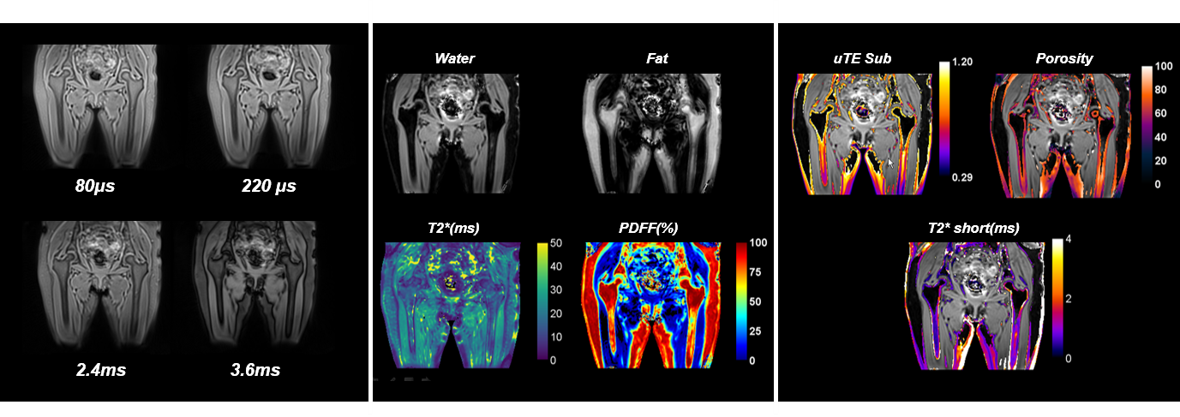
Figure 1: A) Coronal
slices at different echo time extracted from the 3D uTE. B) Parametric
maps from a uTE-CSE acquisition: Fat-water decomposition allowed to reconstruct
adiposity map (PDFF) and long T2 species T2* map. C) uTE
Sub (weighted substraction of long and short echo image) and porosity maps which respectively indicate
average pore size volume and bone porosity, and short T2* map of
cortical bone.
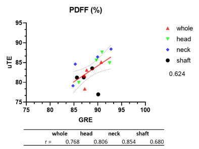
Figure 3: Comparison between PDFF
measurements obtained by uTE and CSE in different hip bone subregions.