1Department of Radiology, Vanderbilt University Institute of Imaging Science, Nashville, TN, United States, 2Department of Radiology, Vanderbilt University Medical Center, Nashville, TN, United States, 3Department of Mechanical Engineering, University of Arkansas, Fayetteville, AR, United States
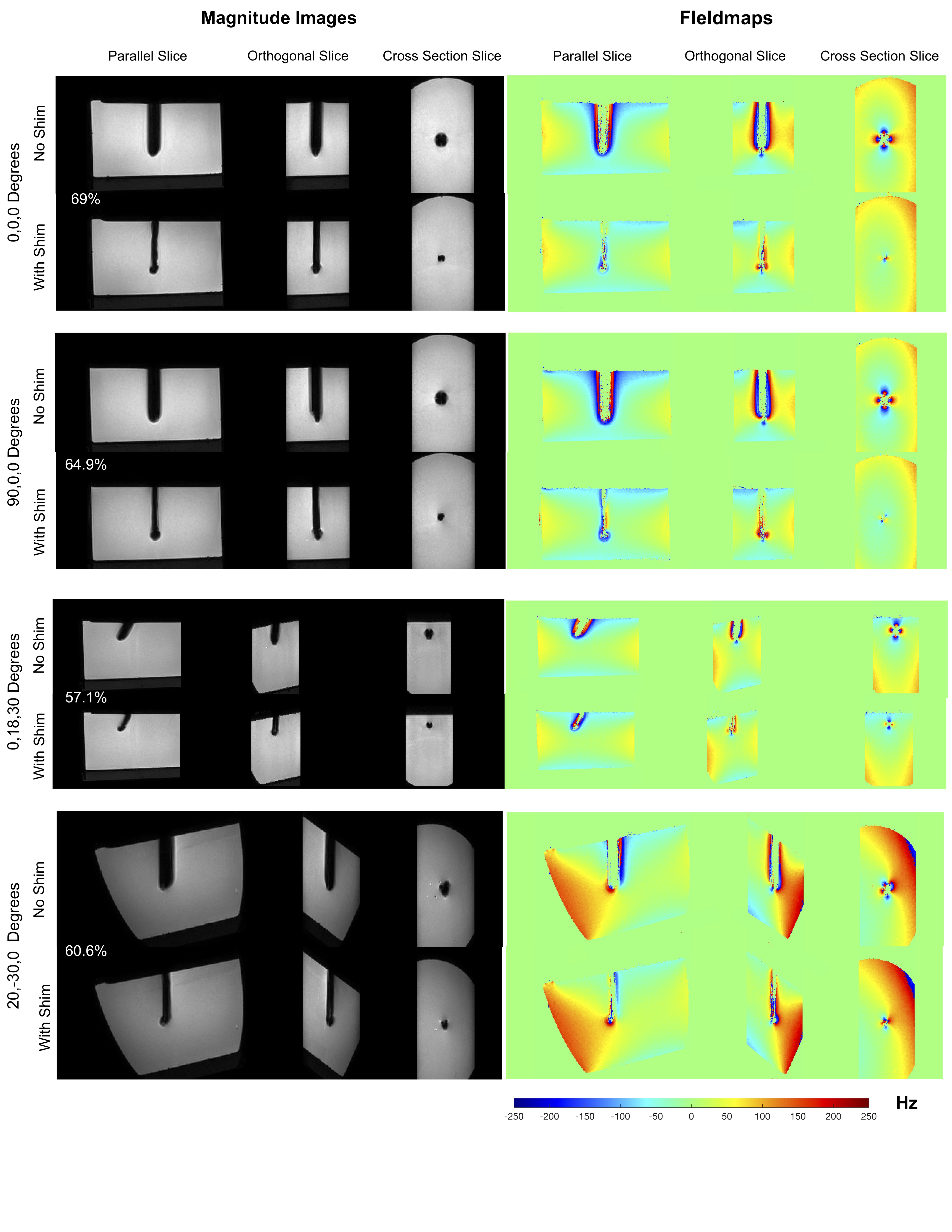
Figure 4
Results of active shimming, 1 x 1 x 1 mm3 3D GRE images and fieldmaps. Excellent recovery of lost signal is achieved in all orientations using pre-estimated shim currents. The width of the signal void approaches the needle width in all cases. Fieldmaps show correction of the underlying ΔB0. Note that the regions closest to the rod with field information in the ‘With Shim’ case have no corresponding field information in the ‘No Shim’ case due to signal loss. The percentage of signal recovered (not accounting for the needle itself) is indicated for all orientations.

Figure 2
Active shimming hardware (a) Shim insert design with bevel and orthogonal slots for shim wires (b) Shim insert inside the titanium needle (c) Needle placed in holder with compartment for electronics (d) Phantoms with guide holes at different angles used for experiments.
