Junghun Cho1, John Lee2, Hongyu An2, Manu S Goyal2, Yi Su3, and Yi Wang1
1Weill Cornell Medicine, New York, NY, United States, 2Washington University School of Medicine, Saint Louis, MO, United States, 3Banner Alzheimers Institute, Phoenix, AZ, United States
1Weill Cornell Medicine, New York, NY, United States, 2Washington University School of Medicine, Saint Louis, MO, United States, 3Banner Alzheimers Institute, Phoenix, AZ, United States
Gradient
echo MRI-based QSM+qBOLD (QQ)-OEF mapping is validated against the reference
standard 15O PET-OEF mapping in healthy adults, with providing substantially equivalent OEF values both globally and regionally.
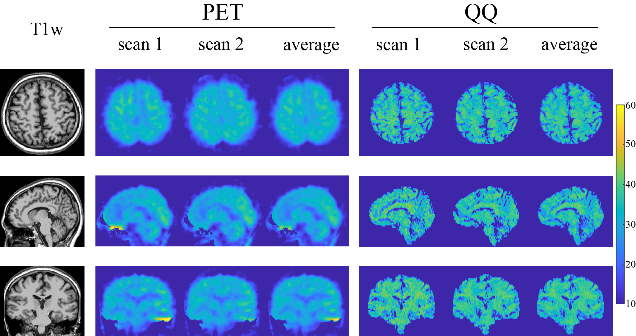
Figure 1. OEF maps from PET and QQ in axial, sagittal, and coronal views in
a subject. Both PET and QQ show uniform OEF maps and good agreement between scans
and methods.
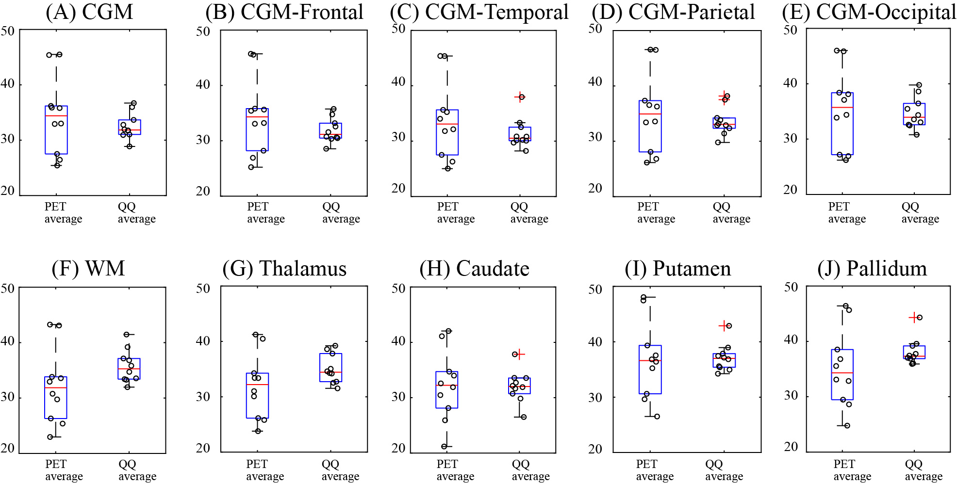
Figure 3. OEF comparison in cortical gray matters (A-E), white matter (F),
and deep gray matters (G-J) among PET and QQ average. No significant difference
was found between PET and QQ (all p-values <0.01, TOST). The unit in y-axis
is %. Red line, blue box, black whisker, and red cross, black
circle indicates median value, interquartile range, the range extending to 1.5
of the interquartile range, outlier beyond the whisker range, and individual
subject value.