Stephan Kaczmarz1, Miriam Reichert1, Moritz Roman Hernandez Petzsche1, Andreas Hock2, Kilian Weiss3, Kim van de Ven4, Christine Preibisch1, Jan Kirschke1, Claus Zimmer1, Makoto Obara5, Michael Helle6, Nico Sollmann1,7, and Hans Liebl1
1School of Medicine, Department of Neuroradiology, Technical University of Munich (TUM), Munich, Germany, 2Philips Healthcare, Horgen, Switzerland, 3Philips Healthcare, Hamburg, Germany, 4Philips Healthcare, Best, Netherlands, 5Philips Japan, Tokyo, Japan, 6University Hospital Schleswig-Holstein Campus Kiel, Kiel, Germany, 7Department of Radiology, University Ulm Medical Center, Ulm, Germany
1School of Medicine, Department of Neuroradiology, Technical University of Munich (TUM), Munich, Germany, 2Philips Healthcare, Horgen, Switzerland, 3Philips Healthcare, Hamburg, Germany, 4Philips Healthcare, Best, Netherlands, 5Philips Japan, Tokyo, Japan, 6University Hospital Schleswig-Holstein Campus Kiel, Kiel, Germany, 7Department of Radiology, University Ulm Medical Center, Ulm, Germany
Non-invasive vascular territory mapping and time-resolved angiography based on super-selective arterial spin labeling (ASL) with automated planning are clinically applicable and highly promising to improve diagnostics in cerebrovascular diseases (CVD)
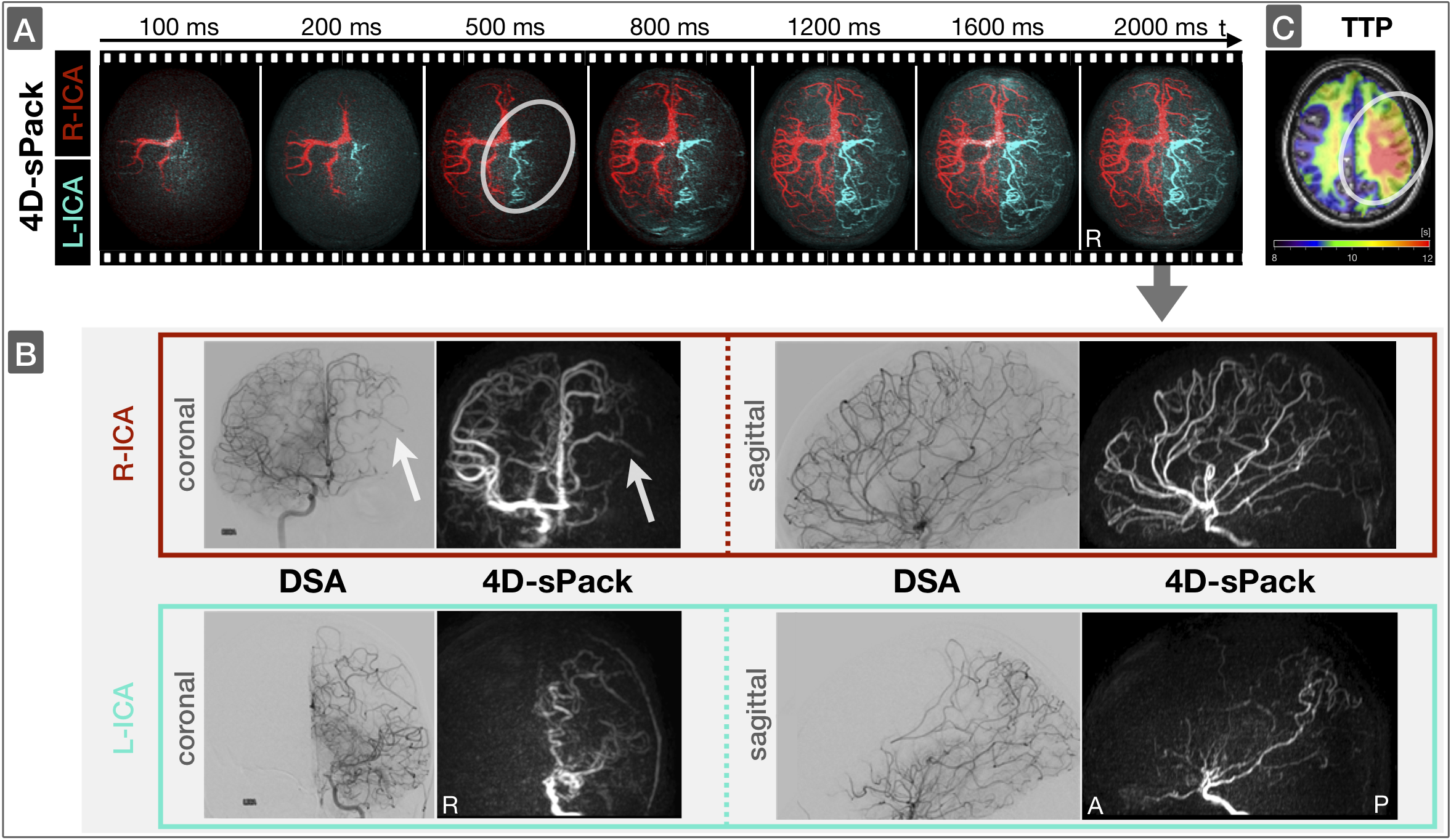
Figure 4: Angiography of a patient with moyamoya
disease. Non-invasive time resolved
angiography by 4D-sPack of R-ICA (red) and L-ICA (cyan) are shown in axial view
with maximum intensity projections (A; top row). The non-invasive angiogram at
t=2000 ms is compared to conventional DSA in coronal and sagittal view for
R-ICA and L-ICA, respectively (B; red & cyan boxes). Note 4D-sPack can even depict small distal collateral
vessels originating from R-ICA in agreement with DSA (B; arrows) as well as
delayed perfusion by L-ICA (A; circles) in agreement with prolonged time-to-peak
(TTP; C).
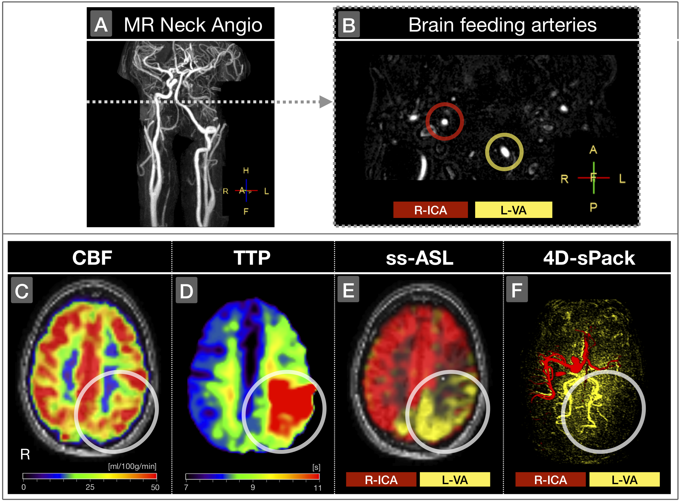
Figure 2: Angiography and perfusion in a patient with
left-ICA dissection, left-M1 occlusion and hypoplastic right-VA. Contrast-enhanced MR neck-angiography (A) in an axial cut shows the cerebral blood supply by R-ICA (red) and L-VA only (yellow; B). CBF is only slightly decreased
in the left posterior hemisphere (circles, C), but with substantially prolonged perfusion delay (D) in this L-VA territory, as depicted by ss-ASL (E). The right-ICA widely
supplies the contralateral hemisphere (E) with takeover of the left anterior
and media territories as demonstrated by 4D-sPack (F).