junxin wang1, yanwei Miao1, and Jiazheng Wang2
1Department of Radiology, the First Affiliated Hospital of Dalian Medical University, Dalian, China, 2Phillips healthcare, dalian, China
1Department of Radiology, the First Affiliated Hospital of Dalian Medical University, Dalian, China, 2Phillips healthcare, dalian, China
This study aimed to exploit the early Wallerian degeneration (WD) along
the corticospinal tract following cerebral ischemic stroke using amide proton transfer
weighted (APTw) and diffusion weighted imaging (DWI).
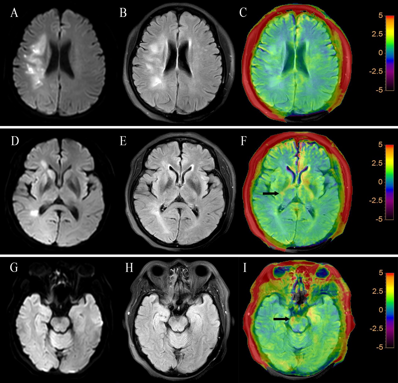
Fig.1 (A,D,G)
DWI, (B,E,H) FLAIR, and (C,F,I) FLAIR+APTw images of an ischemic stroke patient
(female; 56 years old; left limb inactivity for 15 days). DWI images
demonstrated that the patient developed an acute infarction of right frontal
parietal lobe cerebral infarction. The
APTw intensities in regions of the ipsilateral of the ischemic lesion (black
arrow) were higher than the contralateral side in both levels of posterior limb
of internal capsule and cerebral peduncle. DWI
and FLAIR images show no difference between two sides of the two levels.
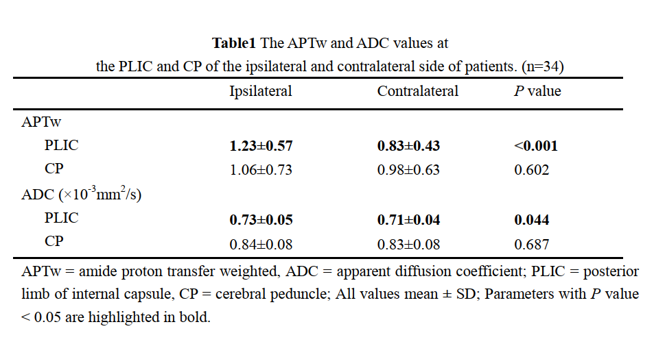
Table1