Zengping Lin1, Tianyao Wang2, Rong Guo3,4, Yudu Li3,4, Yibo Zhao3,4, Tianxiao Zhang1, Jun Liu2, Xin Yu5, Zhi-Pei Liang3,4, and Yao Li1
1School of Biomedical Engineering, Shanghai Jiao Tong University, Shanghai, China, 2Radiology Department, The Fifth People's Hospital of Shanghai, Fudan University, Shanghai, China, 3Beckman Institute for Advanced Science and Technology, University of Illinois at Urbana-Champaign, Urbana, IL, United States, 4Department of Electrical and Computer Engineering, University of Illinois at Urbana-Champaign, Urbana, IL, United States, 5Department of Biomedical Engineering, Case Western Reserve University, Cleveland, OH, United States
1School of Biomedical Engineering, Shanghai Jiao Tong University, Shanghai, China, 2Radiology Department, The Fifth People's Hospital of Shanghai, Fudan University, Shanghai, China, 3Beckman Institute for Advanced Science and Technology, University of Illinois at Urbana-Champaign, Urbana, IL, United States, 4Department of Electrical and Computer Engineering, University of Illinois at Urbana-Champaign, Urbana, IL, United States, 5Department of Biomedical Engineering, Case Western Reserve University, Cleveland, OH, United States
The NAA concentration within the ischemic lesion imaged using fast high-resolution 3D MRSI
decreased in a
time-dependent manner after stroke onset, which might
provide a useful metabolic biomarker for assessment of symptom onset time.
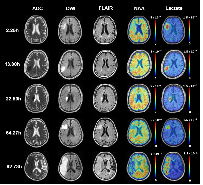
Figure 1. Multimodal images from representative
patients at 2.25 to 92.73 hours after ischemic stroke. The color bar for MRSI shows
NAA or lactate level in institutional units. The NAA signal reduction was
visible within the lesion area and decreased with time.
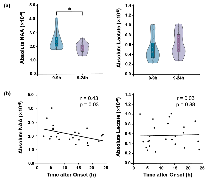
Figure 3. (a) Significant
reduction of NAA was observed for acute stage patients from within to over 9h
window (p < 0.05), but lactate showed no significant difference. (b) A
significant negative correlation between NAA and time after onset was found,
but not for lactate.