Bin Bo1, Tianyao Wang2, Rong Guo3,4, Yudu Li3,4, Yibo Zhao3,4, Tianxiao Zhang1, Zengping Lin1, Ziyu Meng1, Jun Liu2, Xin Yu5, Zhi-Pei Liang3,4, and Yao Li1
1School of Biomedical Engineering, Shanghai Jiao Tong University, Shanghai, China, 2Radiology Department, The Fifth People's Hospital of Shanghai, Fudan University, Shanghai, China, 3Beckman Institute for Advanced Science and Technology, University of Illinois at Urbana-Champaign, Urbana, IL, United States, 4Department of Electrical and Computer Engineering, University of Illinois at Urbana-Champaign, Urbana, IL, United States, 5Department of Biomedical Engineering, Case Western Reserve University, Cleveland, OH, United States
1School of Biomedical Engineering, Shanghai Jiao Tong University, Shanghai, China, 2Radiology Department, The Fifth People's Hospital of Shanghai, Fudan University, Shanghai, China, 3Beckman Institute for Advanced Science and Technology, University of Illinois at Urbana-Champaign, Urbana, IL, United States, 4Department of Electrical and Computer Engineering, University of Illinois at Urbana-Champaign, Urbana, IL, United States, 5Department of Biomedical Engineering, Case Western Reserve University, Cleveland, OH, United States
We
performed fast high-resolution 3D MRSI in a longitudinal cohort of ischemic
stroke patients. Temporal changes of different neurometabolites from acute to
subacute stroke were observed in different regions within the hypoperfused
tissue.
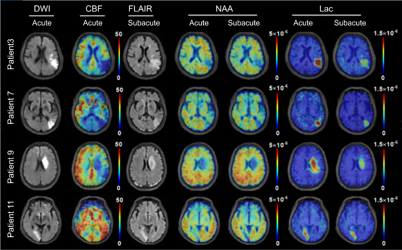
Figure
2. Multimodal images from representative patients at acute and subacute stages after
ischemic stroke. All the images were registered to the structural T1-weighted
images. The color bar for ASL-PWI shows the cerebral blood flow in ml/100g/min.
The color bar for MRSI shows NAA or lactate level in institutional units.
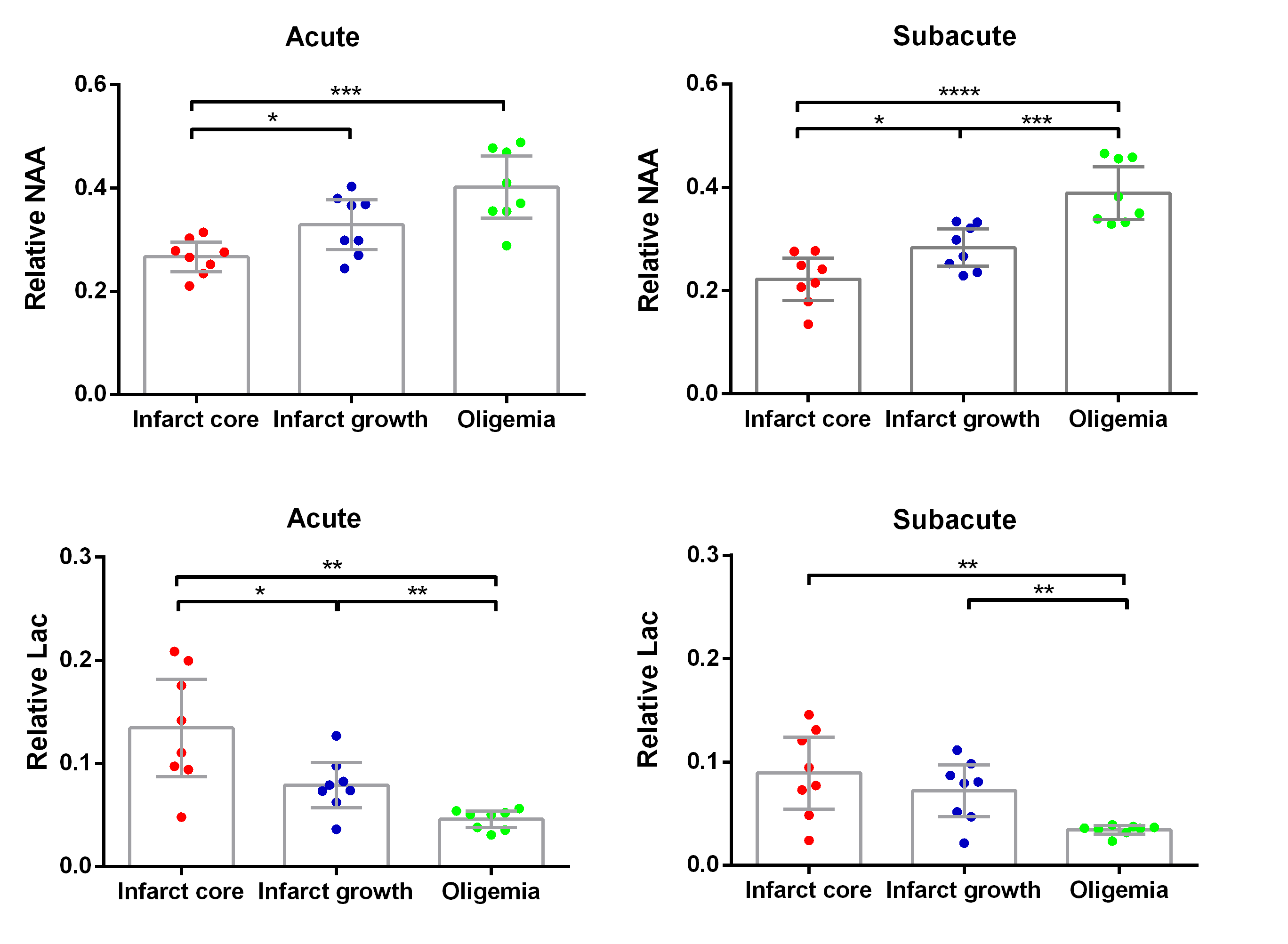
Figure 3. Group comparison of mean
relative NAA and lactate concentrations for different regions of interest in
acute and subacute stroke patients, respectively (Error bars, 95% confidence
interval). *p < 0.05, **p < 0.01.