Di Wu1, Shun Zhang1, and Weiyin Vivian Liu2
1Radiology, Tongji Hospital, Tongji Medical College, Huazhong University of Science and Technology, Wuhan, Hubei, China, 2MR Research, GE Healthcare, Beijing, China, Beijing, China
1Radiology, Tongji Hospital, Tongji Medical College, Huazhong University of Science and Technology, Wuhan, Hubei, China, 2MR Research, GE Healthcare, Beijing, China, Beijing, China
Difference of CBF was found between patients with and
without penumbra. The OEF and nonblood susceptibility
of tissue in the penumbra were significantly different from ischemic
core. OEF in the diffusion lesion was correlated with
clinical severity and continuously decreased with time.
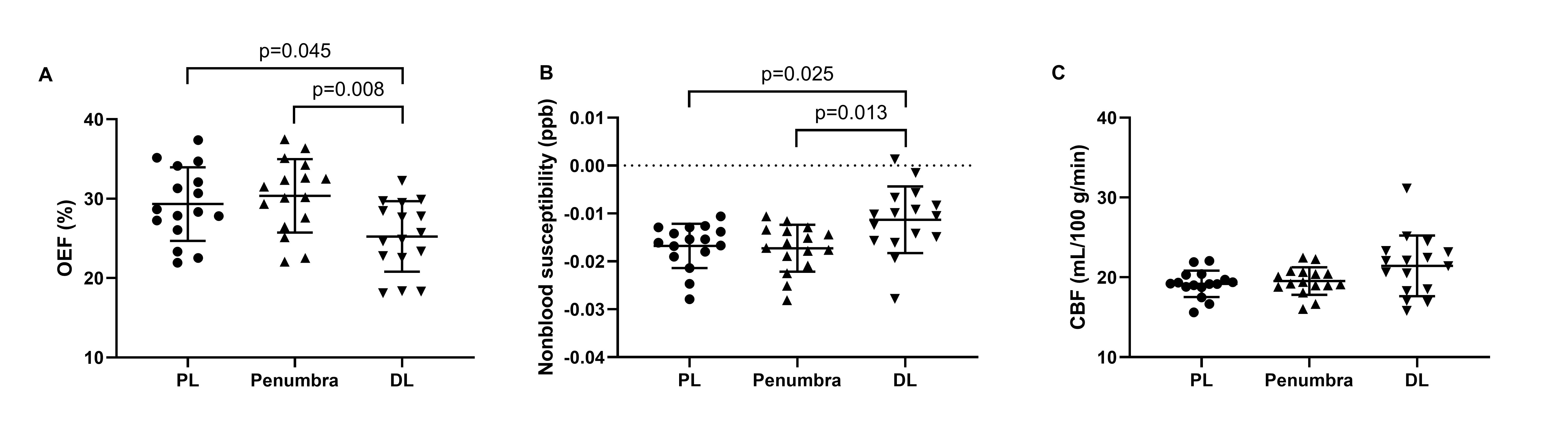
Figure 2. Oxygen
extraction fraction (OEF, A), nonblood susceptibility (B) and cerebral blood
flow (CBF, C) in the perfusion lesion (PL), penumbra and diffusion lesion (DL)
of the mismatched group. Significant differences of OEF and nonblood
susceptibility were found between DL and PL and between DL and penumbra. There
were no significant differences of CBF among PL, DL and penumbra.
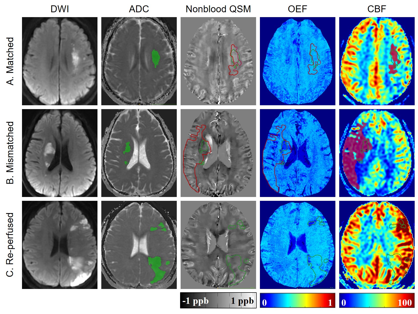
Figure 1.
Representative images of three patients in the corresponding groups classified
by DEFUSE 3 criteria. (A) A 64-year-old female with the volume of diffusion
lesion (shown in green on ADC maps) matched with that of perfusion lesion
(shown in red on CBF maps). (B) A 50-year-old male whose perfusion lesion was
rather larger than the diffusion lesion. (C) A 33-year-old female with re-perfused
left hemisphere.