Tobias Speidel1, Dagmar Bertsche1, Patrick Metze1, Kilian Stumpf1, Wolfgang Rottbauer1, and Volker Rasche1
1Internal Medicine II, Ulm University Hospital, Ulm, Germany
1Internal Medicine II, Ulm University Hospital, Ulm, Germany
Qualitative 3D isotropic heart images were acquired within 250 heartbeats (ECG gating), using specifically generated k-space interleaves with low-coherent and low-discrepancy sampling properties, based on a generalized form of the previously introduced Seiffert-Spirals.
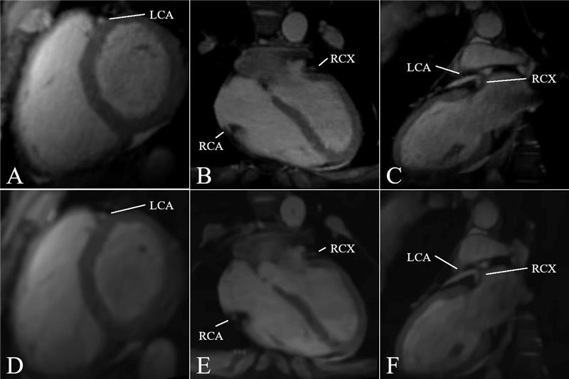
Figure 2: Axial, coronal and sagittal anatomical planes of the 3D $$$\zeta$$$-based acquisition. A,B,C) was reconstructed using the entire dataset of 1,000 heart beats while D,E,F) consists of the first 250 heart beats.
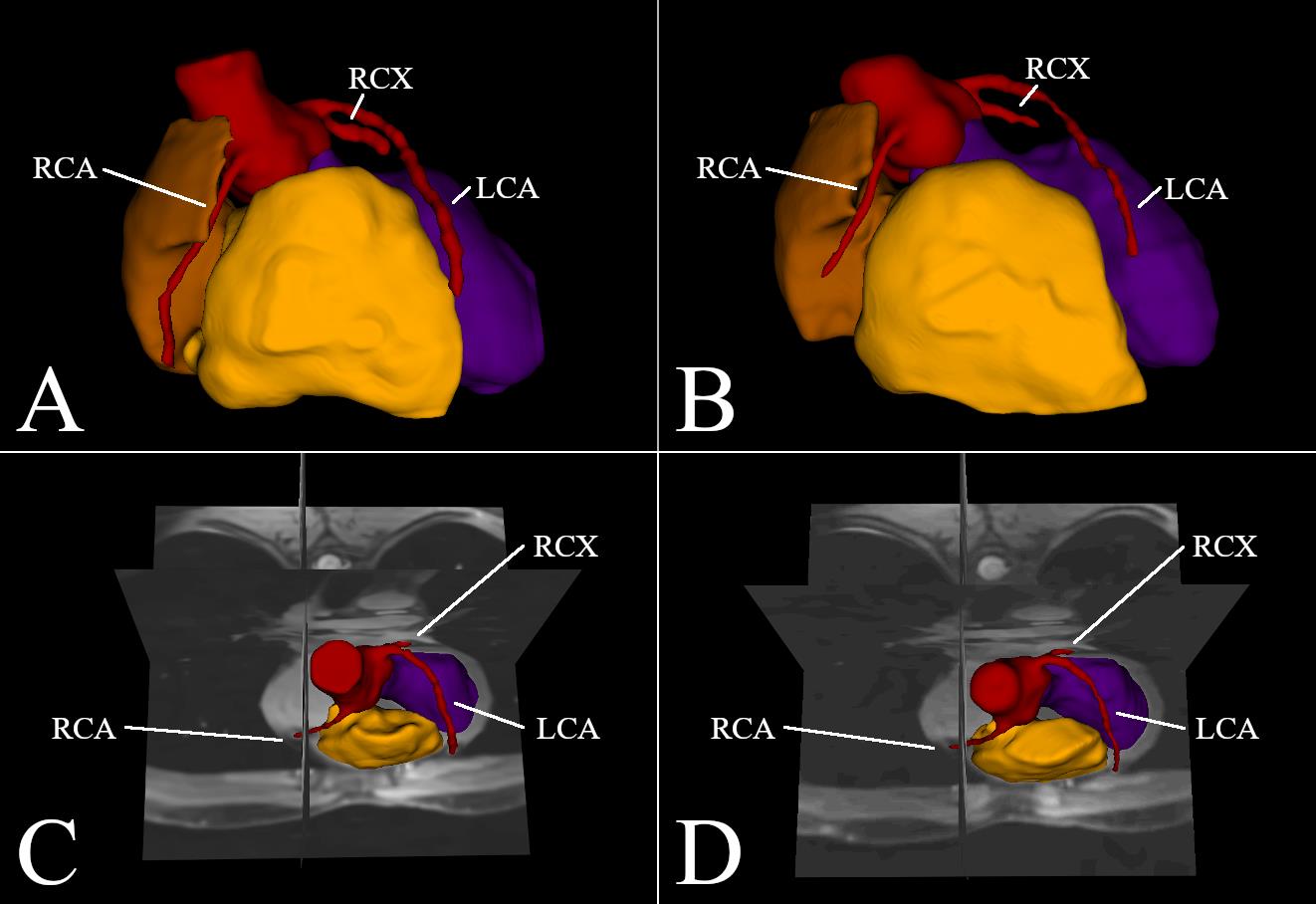
Figure 5: 3D MRI cross-sections in addition to segmented LCA, RCA and RCX as well as a segmented left ventricle, right ventricle and right atrium. A,C) was reconstructed using the entire dataset of 1,000 heart beats while B,D) is based on the first 250 heart beats.