Amir Seginer1, Edna Furman-Haran2,3, Ilan Goldberg4, and Rita Schmidt3,5
1Siemens Healthcare, Rosh Ha'ayin, Israel, 2Life Sciences Core Facilities, Weizmann Institute of Science, Rehovot, Israel, 3The Azrieli National Institute for Human Brain Imaging and Research, Weizmann Institute of Science, Rehovot, Israel, 4Deparment of Neurology, Wolfson medical center, Holon, Israel, 5Neurobiology Department, Weizmann Institute of Science, Rehovot, Israel
1Siemens Healthcare, Rosh Ha'ayin, Israel, 2Life Sciences Core Facilities, Weizmann Institute of Science, Rehovot, Israel, 3The Azrieli National Institute for Human Brain Imaging and Research, Weizmann Institute of Science, Rehovot, Israel, 4Deparment of Neurology, Wolfson medical center, Holon, Israel, 5Neurobiology Department, Weizmann Institute of Science, Rehovot, Israel
- SAR may limit volume coverage and min. TR for fMRI at 7T.
- Removing the fat suppression pulse from GRE-EPI greatly reduces SAR.
- To compensate, an SMS-like reconstruction separates lipid and water images.
- Simulations, phantom experiments, and fMRI experiments support the method.
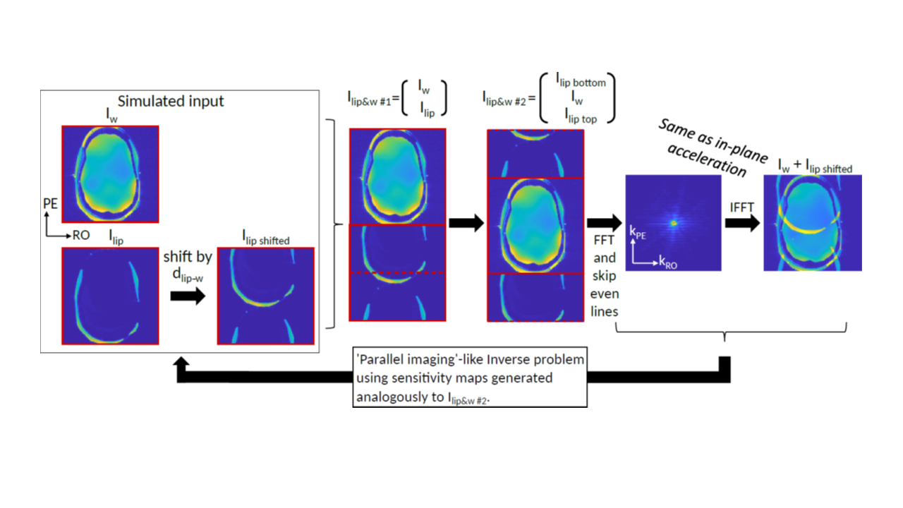
Figure 1: Extended
formulation unifying three parallel imaging aspects:
(i) in-plane PE acceleration, (ii) SMS acceleration and (iii)
lipid-water separation. The steps show how an EPI image (far right) is
equivalent to an SMS of water and lipid “slices” (Iw and Ilip) and to an in-plane PE acceleration (Ilip&w #2 vs. Iw+Ilip shifted). The formulation can be extended
to any number of slices and fat/water images.
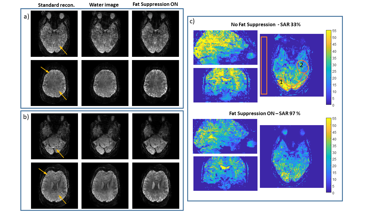
Figure 5: Resting-state fMRI. a),b) Examples of simultaneously acquired slices. Left to right: standard reconstruction, water image after lipid separation, and a fat suppressed image. Arrows mark artifacts from the lipids. (c) tSNR on three orthogonal planes. SNR and tSNR in main text where estimated from marked regions. SAR was 33% without fat-suppression and 97% with fat-suppression (reference amplitude of 240 V for a 1 ms 180° hard pulse). Scan parameters: in-plane accelerations ×3, SMS ×2, FOV=220×220 mm2, resolution = 1.7×1.7 mm2, slice thickness = 1.7 mm, TR/TE = 1500/22 ms.