Jaeyong Yu1,2, Seulki Yoo1,2, Jae-Kyun Ryu3, Seung-Kyun Lee1,2,4, and Jang-Yeon Park1,2,3
1Department of Biomedical Engineering, Sungkyunkwan University, Suwon, Korea, Republic of, 2Department of Intelligent Precision Healthcare Convergence, Sungkyunkwan University, Suwon, Korea, Republic of, 3Biomedical Institute for Convergence at SKKU, Sungkyunkwan University, Suwon, Korea, Republic of, 4Center for Neuroscience Imaging Research, Institute for Basic Science (IBS), Suwon, Korea, Republic of
1Department of Biomedical Engineering, Sungkyunkwan University, Suwon, Korea, Republic of, 2Department of Intelligent Precision Healthcare Convergence, Sungkyunkwan University, Suwon, Korea, Republic of, 3Biomedical Institute for Convergence at SKKU, Sungkyunkwan University, Suwon, Korea, Republic of, 4Center for Neuroscience Imaging Research, Institute for Basic Science (IBS), Suwon, Korea, Republic of
We
proposed a new method for ERASE sequence with head-tilted scan to
improve B0 inhomogeneity in comparison with gradient-echo
EPI.
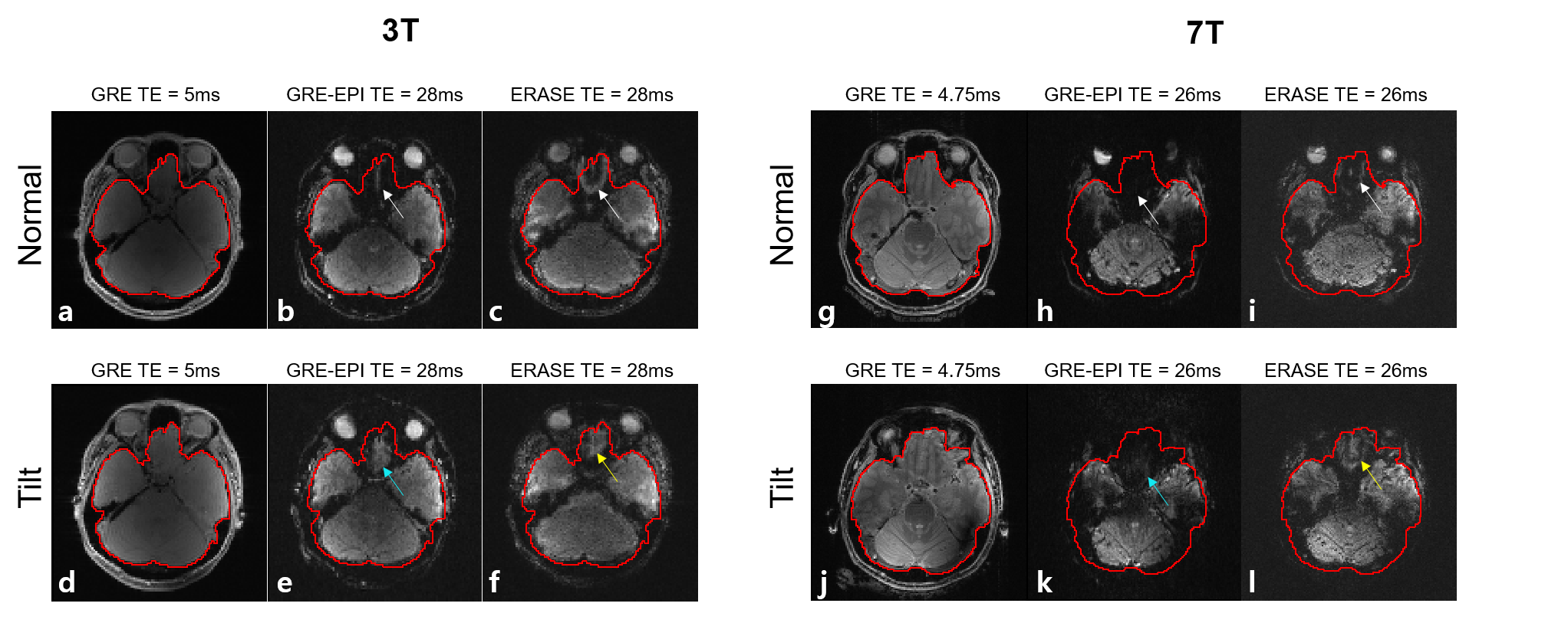
Figure 3. Axial views of the
reference GRE magnitude, EPI and ERASE images for a single subject. The solid
red lines indicate the manually drawn masks as a guide for
comparison of image quality. Signal loss and image degradation in prefrontal
cortex of the normal-orientation scans (white arrows) are reduced in
tilted scans (blue arrows and yellow arrows).
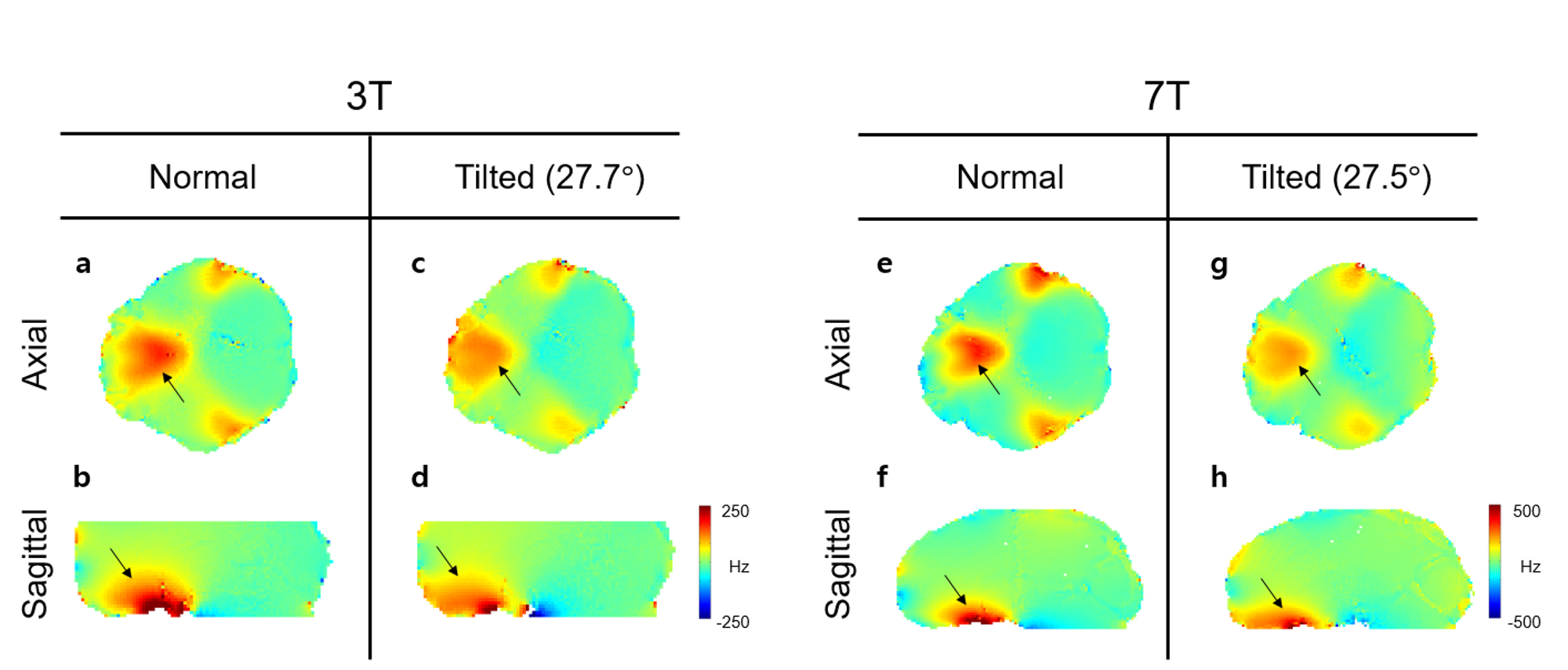
Figure 2. Measured B0 maps at normal and tilted head
orientations at 3T and 7T. Black arrows indicate B0 inhomogeneity near the prefrontal
region. In head-tilted scan (c-d, g-h), B0 field distribution is spread more indicating that
local gradient is smaller
than the normal orientation scan (a-b, e-f).