Johannes Fischer1, Ali Caglar Özen1,2, Matthias Echternach3, Louisa Traser4, Bernhard Richter4, and Michael Bock1
1Dept.of Radiology, Medical Physics, Medical Center University of Freiburg, Faculty of Medicine, University of Freiburg, Freiburg, Germany, 2German Consortium for Translational Cancer Research Freiburg Site, German Cancer Research Center (DKFZ), Heidelberg, Germany, 3Division of Phoniatrics and Pediatric Audiology, Department of Otorhinolaryngology, Head and Neck Surgery, Ludwig-Maximilians-University, Munich, Germany, 4Institute of Musicians' Medicine, Freiburg University Medical Center, Germany Faculty of Medicine, University of Freiburg, Freiburg, Germany
1Dept.of Radiology, Medical Physics, Medical Center University of Freiburg, Faculty of Medicine, University of Freiburg, Freiburg, Germany, 2German Consortium for Translational Cancer Research Freiburg Site, German Cancer Research Center (DKFZ), Heidelberg, Germany, 3Division of Phoniatrics and Pediatric Audiology, Department of Otorhinolaryngology, Head and Neck Surgery, Ludwig-Maximilians-University, Munich, Germany, 4Institute of Musicians' Medicine, Freiburg University Medical Center, Germany Faculty of Medicine, University of Freiburg, Freiburg, Germany
Single point imaging with rapid encoding
(SPIRE) is used image the vocal fold oscillations in the coronal plane
and readout in AP direction allows for the reconstruction in multiple slices.
Electroglottography and MR navigators are used to gate and motion-correct data
prior to reconstruction.
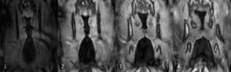
Figure 4: Vocal
fold oscillation in the coronal plane reconstructed into 4 slices and 12 Frames
resulting in a temporal resolution Δt = 890μs (f0 = 92Hz). Although motion of
the vocal folds is visible, no full contact can be observed. The vocal fold
oscillations cause the EGG electrodes to vibrate on the skin, which can be seen
on the edges of each frame.
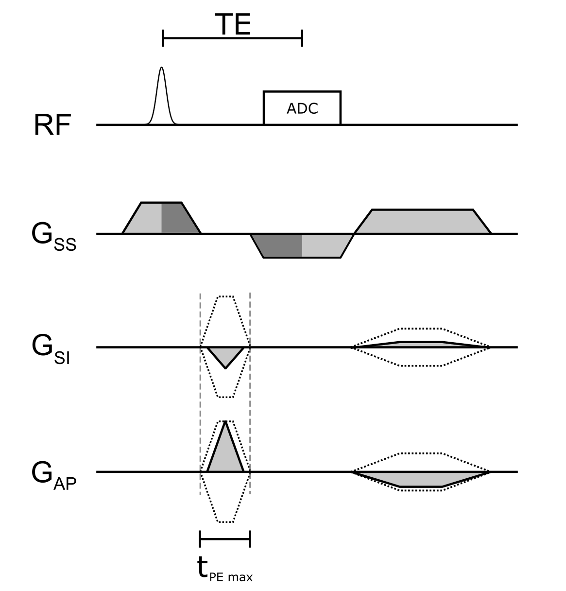
Figure 1: Sequence
diagram of the 2.5D SPIRE method. Rapidly switched phase encoding gradients
immediately follow $$$G_\mathrm{SS}$$$ and slice rephasing is performed simultaneously with
the frequency encoding in slice direction (dark grey areas indicate equal
gradient moments). PE gradients are rewound and a spoiler is applied with
opposite sign to the readout gradient to reduce switching when ramping to $$$G_\mathrm{SS}$$$
at the beginning of the next acquisition block. Dotted lines indicate the
gradient shapes for the largest phase encoding moments.