Serhat Ilbey1, Johannes Fischer1, Michael Bock1, and Ali Caglar Ozen1,2
1Dept. of Radiology, Medical Physics, Medical Center University of Freiburg, Faculty of Medicine, University of Freiburg, Freiburg, Germany, 2German Consortium for Translational Cancer Research Partner Site Freiburg, German Cancer Research Center (DKFZ), Heidelberg, Germany
1Dept. of Radiology, Medical Physics, Medical Center University of Freiburg, Faculty of Medicine, University of Freiburg, Freiburg, Germany, 2German Consortium for Translational Cancer Research Partner Site Freiburg, German Cancer Research Center (DKFZ), Heidelberg, Germany
csPETRA
enables 3D imaging with isotropic sub-millimeter resolution within only a few minutes,
e.g., for (0.5 mm)3 voxel size with (20 cm)3 field of
view, which is demonstrated with different acceleration factors for high-resolution
imaging of the knee.
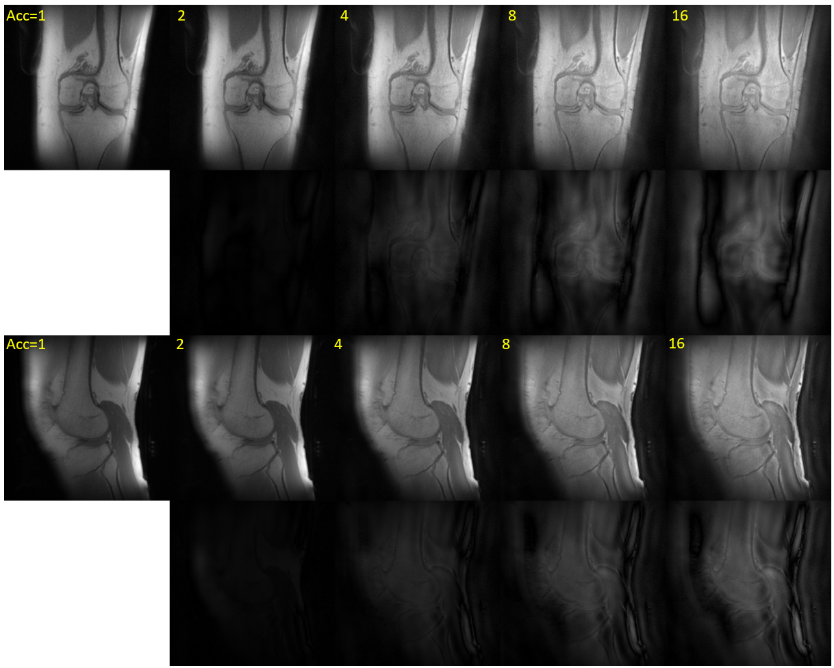
Fig. 3: Coronal (first row) and sagittal view (third row) of PETRA (Acc=1) and csPETRA (Acc>1) images. Yellow numbers show the Acc factor used for the SPI acquisition. Second and fourth rows show difference images subtracted with Acc=1 images (far left images) of coronal and sagittal views, respectively.
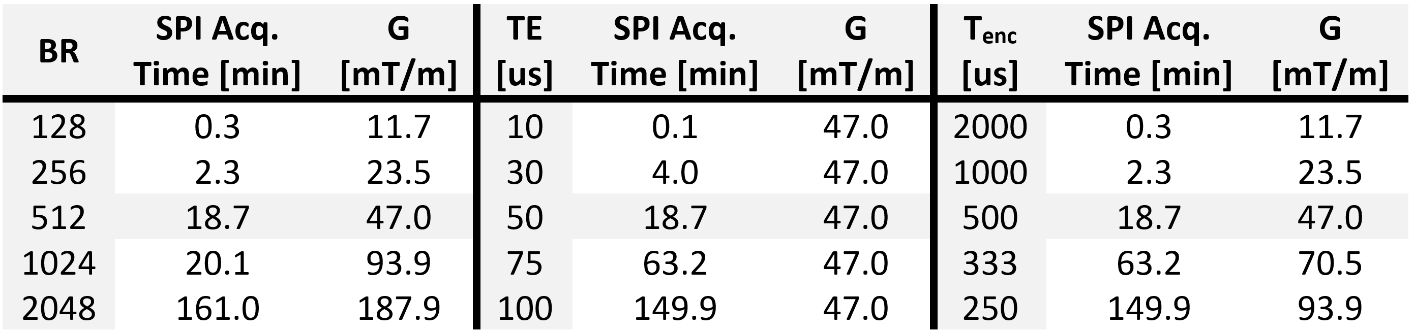
Table 1: Examples of SPI acquisition times w.r.t. the parameters $$$\text{BR}$$$, $$$\text{TE}$$$, and $$$T_\text{enc}$$$ with the corresponding maximum gradient amplitude (|G|). Default parameters were: $$$\text{BR}$$$=512, $$$\text{TE}$$$=50 µs, $$$T_\text{enc}$$$=500 us, TR=2 ms, when they were not used as the sweep parameter.