Ka-Loh Li1, Daniel Lewis2, David J Coope3, Federico Roncaroli3, Omar N Pathmanaban2, Andrew T King2, Sha Zhao1, Erjon Agushi1, Alan Jackson1, Timothy Cootes1, and Xiaoping Zhu1
1Division of Informatics, Imaging and Data Sciences, The University of Manchester, Manchester, United Kingdom, 2Department of Neurosurgery, Salford Royal NHS Foundation Trust, Manchester, United Kingdom, 3Division of Neuroscience and Experimental Psychology, The University of Manchester, Manchester, United Kingdom
1Division of Informatics, Imaging and Data Sciences, The University of Manchester, Manchester, United Kingdom, 2Department of Neurosurgery, Salford Royal NHS Foundation Trust, Manchester, United Kingdom, 3Division of Neuroscience and Experimental Psychology, The University of Manchester, Manchester, United Kingdom
A new dual-temporal resolution DCE-MRI analysis technique (LEGATOS) combined with a single-injection low-dose interleaved protocol permits the derivation of accurate, tissue-validated high-spatial resolution kinetic parameter estimates following a single low contrast agent dose.
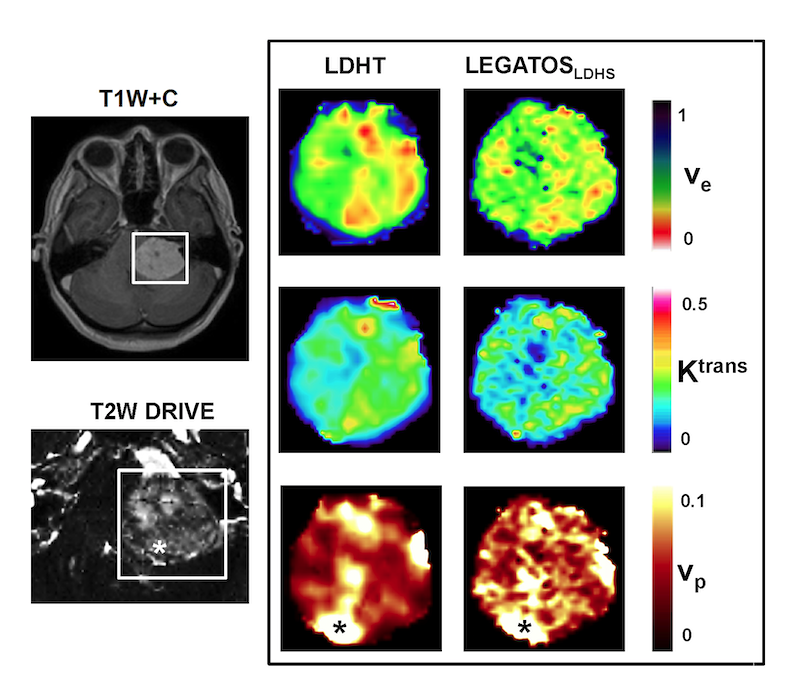
Figure 3: Representative images from a patient with a large left sided sporadic VS imaged using a single low-dose (LD) interleaved high-temporal (HT) (frame duration of Dt = 1.46 s) and high-spatial resolution (Dt = 6.04 s) DCE-MRI acquisition on a 1.5 T Philips scanner. Kinetic maps derived using the HT segments alone (LDHT, left column) or the LEGATOS method are shown. Note the increased vascularity around the tumor capsule, visible on both the high-spatial T2W DRIVE acquisition (voxel size = 0.5 x 0.5 x 0.5mm3) and LEGATOS derived vp parameter maps (*).
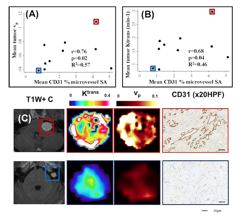
Figure 4: Comparison of LEGATOS derived vp and Ktrans estimates from the in vivo single-injection low-dose DTR Study against tissue derived parameters
(A) Inter-tumor scatterplot comparison of LEGATOS derived mean tumor vp estimates against mean CD31 % microvessel surface area (SA).
(B) Inter-tumor scatterplot analysis of LEGATOS derived mean tumor Ktrans (min-1) against mean CD31 % microvessel surface area (SA).
(C) Representative imaging and histology from a patient with a growing highly vascular VS (top row) and a comparatively less vascular static VS (bottom row) are shown.