Jenny Yang1, Mani Salarian2, Hua Yang3, Shanshan Tan4, Oluwatosin Y Ibhagui4, Jingjuan Qiao4, Zongxiang Gui4, and Hans E Grossniklaus3
1Chemistry, Georgia State University, Atlatna, GA, United States, 2Chemistry, Georgia State University, Atlanta, GA, United States, 3Emory University, Atlanta, GA, United States, 4Georgia State University, Atlanta, GA, United States
1Chemistry, Georgia State University, Atlatna, GA, United States, 2Chemistry, Georgia State University, Atlanta, GA, United States, 3Emory University, Atlanta, GA, United States, 4Georgia State University, Atlanta, GA, United States
We anticipate that pMRI will demonstrate
significantly-improved imaging sensitivity and accuracy and will enable
detection of liver metastasis at a much earlier stage, potentially leading to
improved treatment responses.
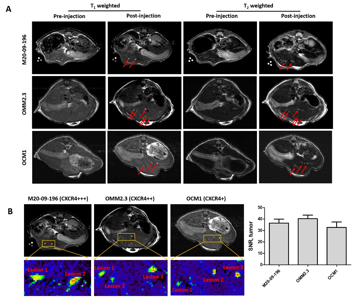
Figure 4. . MR images of metastatic
mice models with ProCA32.CXCR4 administration. A. Comparison of MRI images of
metastatic mice models including M20-09-196, OMM2.3, and OCM1 before and after
administration of ProCA32.CXCR4. B. Zoom-in view of the metastases from M20-09-196,
OMM2.3, and OCM1 mouse models; MRI signal-noise-ratio (SNR) of metastases
following the ProCA32.CXCR4 administration.
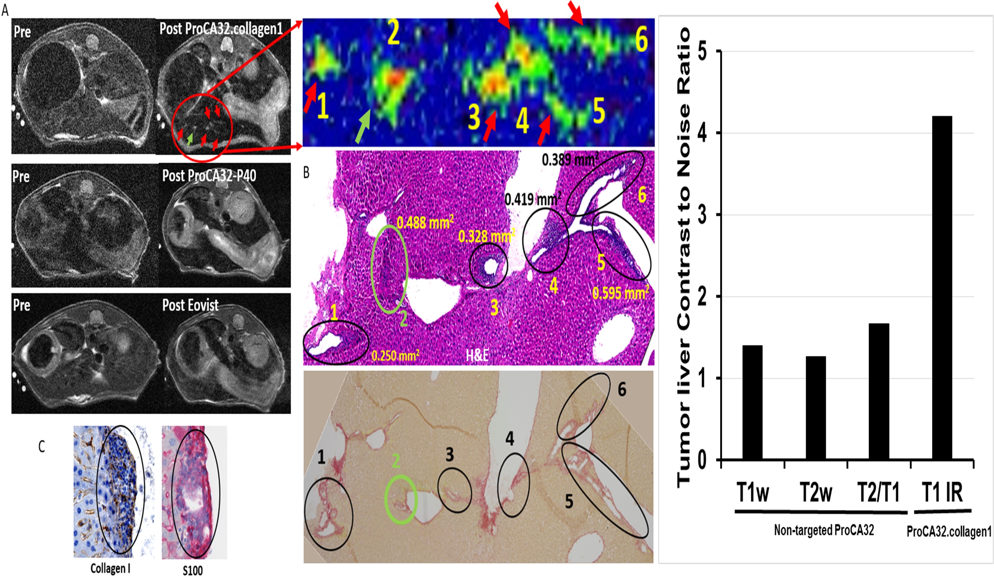
Figure 2. A Detection of UM
liver metastasis using ProCA32.collagen (top), non-targted ProCA32 (middle) and
Eovist (bottom) at 7T. Detected liver
metastasis are verified by H&E and Sirius red staining (B) and UM markers HMB45 and S100 staining (C). The tumor liver contrast to noise ratio is
further increased by inversion recovery pulse sequence showed in the figure on right.