Nathaniel Kim1, Arsen Mamakhantan1, Kristin Granlund1, Elisa de Stanchina2, Manish Shah3, Lewis Cantley4, and Kayvan R. Keshari1
1Department of Radiology, Memorial Sloan Kettering Cancer Center, New York, NY, United States, 2Antitumor Assessment Core Facility, Molecular Pharmacology Program, Memorial Sloan Kettering Cancer Center, New York, NY, United States, 3Weill Cornell Medicine, New York-Presbyterian Hospital, New York, NY, United States, 4Meyer Cancer Center, Department of Medicine, Well Cornell Medical College, New York, NY, United States
1Department of Radiology, Memorial Sloan Kettering Cancer Center, New York, NY, United States, 2Antitumor Assessment Core Facility, Molecular Pharmacology Program, Memorial Sloan Kettering Cancer Center, New York, NY, United States, 3Weill Cornell Medicine, New York-Presbyterian Hospital, New York, NY, United States, 4Meyer Cancer Center, Department of Medicine, Well Cornell Medical College, New York, NY, United States
Hyperpolarized dehydroascorbic acid was used to measure changes in redox in KRAS and BRCA mutant PDX models of pancreatic cancer. Increasing the hyperpolarized lifetime via D2O solvation in awake mouse injections allowed for changes in oxidative stress to be measured upon ascorbate therapy.
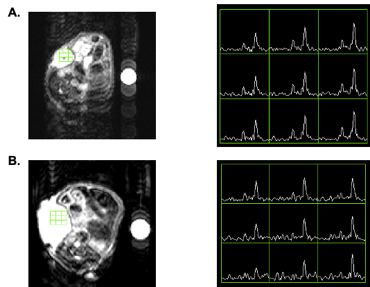
Figure 3. Representative HP 13C MRS of (A) vehicle and (B) ascorbate-treated KRAS PDX model of pancreatic cancer. Metabolic conversion of DHA to ascorbate in pancreatic cancer PDXs changes after 1 week treatment of twice daily ascorbate.

Figure 2. Tumor growth curves for patient derived xenograft models of KRAS and BRCA driven cancer. After 2 weeks of tumor implantation, mice were randomly divided into two groups. One group was treated with freshly prepared vitamin C in 400 μL of PBS (4 g/kg) twice a day via IP injection (KRAS, n= 7; BRCA, n = 7). Control group mice were treated with PBS using the same twice a day dose (KRAS, n = 7; BRCA, n = 7). Tumor growth change for the KRAS PDX tumors is significant after 21 days and significant after 4 days for the BRCA PDX tumors.