Lawrence Lechuga1, Sean B Fain1,2,3,4, Christian M Capitini3,4,5, and Matthew H Forsberg5
1Medical Physics, University of Wisconsin, Madison, Madison, WI, United States, 2Radiology, University of Wisconsin, Madison, Madison, WI, United States, 3Biomedical Engineering, University of Wisconsin, Madison, Madison, WI, United States, 4Carbone Cancer Center, University of Wisconsin, Madison, Madison, WI, United States, 5Pediatrics, University of Wisconsin, Madison, Madison, WI, United States
1Medical Physics, University of Wisconsin, Madison, Madison, WI, United States, 2Radiology, University of Wisconsin, Madison, Madison, WI, United States, 3Biomedical Engineering, University of Wisconsin, Madison, Madison, WI, United States, 4Carbone Cancer Center, University of Wisconsin, Madison, Madison, WI, United States, 5Pediatrics, University of Wisconsin, Madison, Madison, WI, United States
PFPE labeled NK cells were detected and quantified in 3 lymphoma bearing mice out to 6 days post injection by 19F MRI. Quantification indicates that 87% and 70% were detectible at days 0 and 6. Postmortem flow cytometry verified that NK cells maintain label and viability out to 6 days post injection.
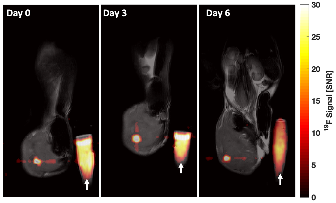
Representative mouse composite magnitude images on Day 0, 3, and 6 after intratumoral injection of 5.3 x 10⁵ NK cells into syngeneic EL4 lymphomas. Images were scaled against their own noise to place into units of pixel SNR. The 19F signal mitigating from labeled cells is detectible within the tumor in all three images. White arrow indicates the PFPE reference vial of known spin density.
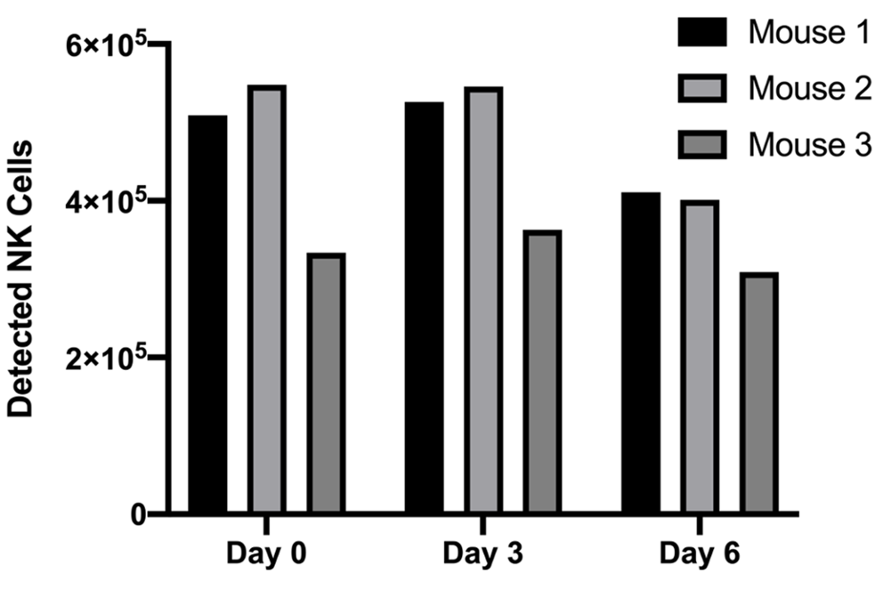
In vivo NK cell quantification results indicate the calculated number of PFPE-red labeled GFP+ NK cells within the tumor volume of each mouse. Difference in time points are not statistically significant.