Vanessa L. Franke1, Justyna Platek1, Philip S. Boyd1, Stephanie Laier2, Karin Mueller-Decker2, Andrey Glinka3, Mark E. Ladd1, Steffen Goerke1, Peter Bachert1, and Andreas Korzowski1
1Division of Medical Physics in Radiology, German Cancer Research Center (DKFZ), Heidelberg, Germany, 2Center for Preclinical Research, Core Facility Tumor Models, German Cancer Research Center (DKFZ), Heidelberg, Germany, 3Division of Molecular Embryology, German Cancer Research Center (DKFZ), Heidelberg, Germany
1Division of Medical Physics in Radiology, German Cancer Research Center (DKFZ), Heidelberg, Germany, 2Center for Preclinical Research, Core Facility Tumor Models, German Cancer Research Center (DKFZ), Heidelberg, Germany, 3Division of Molecular Embryology, German Cancer Research Center (DKFZ), Heidelberg, Germany
31P
MRSI with large spatial coverage in mice at B0=9.4T with a spatial
resolution of (2.5x2.5x7.5)mm³ obtained in 50 minutes is feasible and enables
the quantification of signals in diseased tissue, while maintaining the
separation to healthy tissue
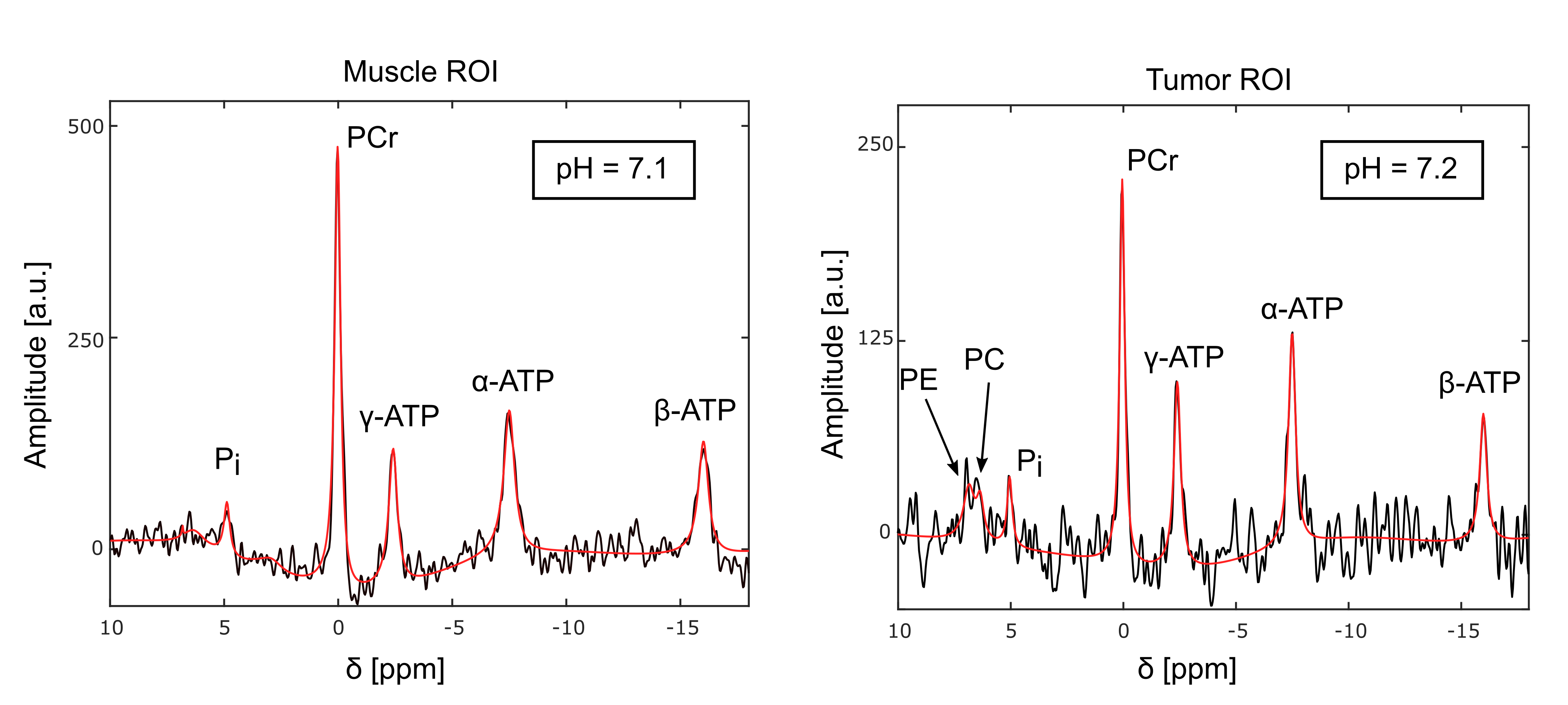
Figure 4: Summed 31P spectra from
the muscle (left) and tumor ROI (right) with the corresponding fitted signal
(red line) acquired with protocol 2 (Fig.3). Acquisition parameters: TR=300ms, α=45°, Δf=6060 Hz, 1024
time points, postprocessing with a 40-Hz Gaussian filter in time domain.
The following resonances were
resolved: PCr, ATP, Pi, PC, and Phosphoethanolamine (PE). Note the
different scales of the y-axis in both spectra.
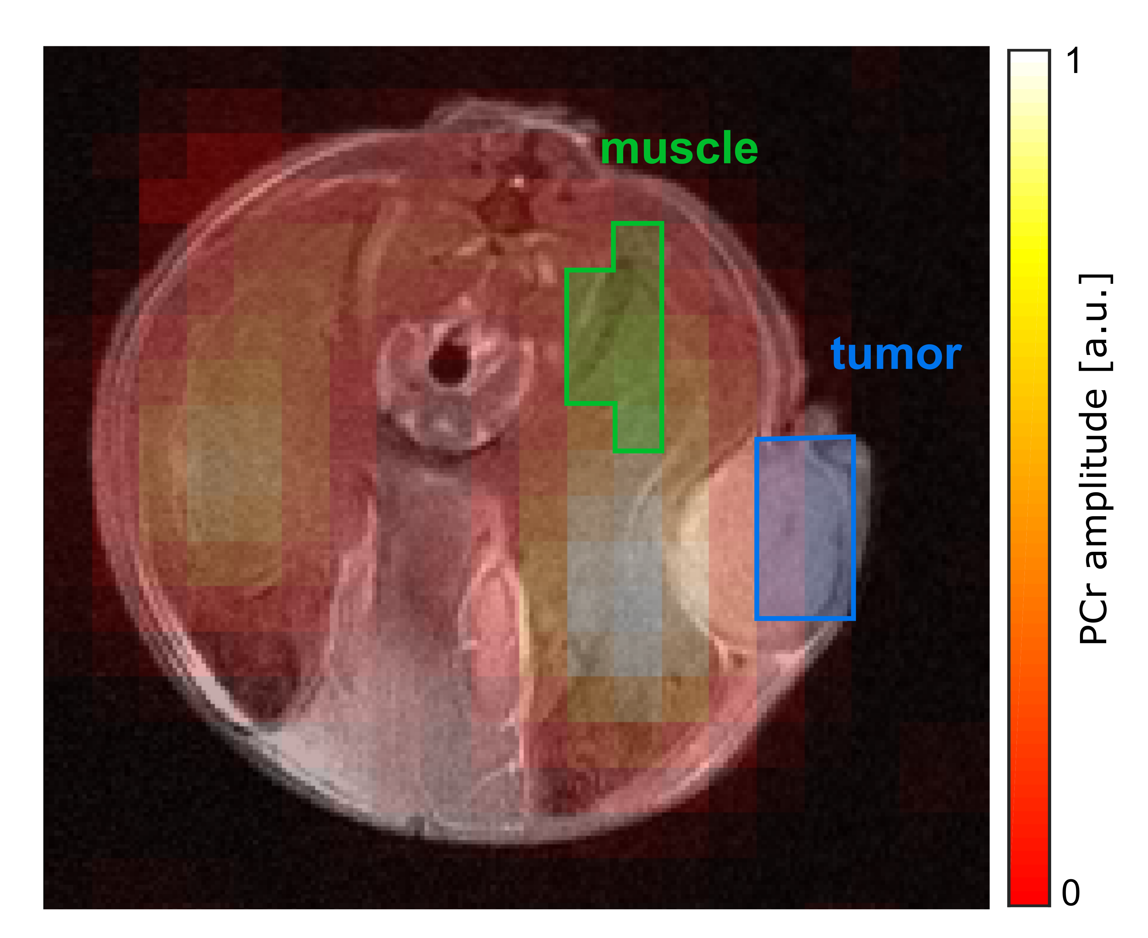
Figure 3: Transversal map of
the fitted PCr amplitude overlaid on the morphological 1H image in
the slice covering the tumor. Data was acquired with protocol 2: matrix size=10x10,
FOV=(25x25)mm², Hamming-weighted k-space averaging with 400 central averages, postprocessing with zerofilling-factor 2. The tumor ROI (blue) was
drawn on the marginal part of the tumor to reduce signal contamination from muscle. The muscle ROI (green) includes the same number of voxels as the
tumor ROI.