Emma L. Reeves1, Jin Li1,2, Konstantinos Zormpas-Petridis1, Jessica K. R. Boult1, James Sullivan1,3, Craig Cummings1, Barbara Blouw4, David Kang4, Ralph Sinkus5, Yann Jamin1, Jeffrey C. Bamber1, and Simon P. Robinson1
1Radiotherapy & Imaging, Institute of Cancer Research, London, United Kingdom, 2Institutes of Brain Science, Fudan University, Shanghai, China, 3Royal Marsden NHS Foundation Trust, Sutton, United Kingdom, 4Halozyme Therapeutics, San Diego, CA, United States, 5Division of Imaging Sciences and Biomedical Engineering, King's Health Partners, St Thomas's Hospital, London, United Kingdom
1Radiotherapy & Imaging, Institute of Cancer Research, London, United Kingdom, 2Institutes of Brain Science, Fudan University, Shanghai, China, 3Royal Marsden NHS Foundation Trust, Sutton, United Kingdom, 4Halozyme Therapeutics, San Diego, CA, United States, 5Division of Imaging Sciences and Biomedical Engineering, King's Health Partners, St Thomas's Hospital, London, United Kingdom
MRE revealed a ~80% increase in MDA-MB-231 LM2-4 tumour viscoelasticity following hyaluronan (HA) degradation by PEGPH20. However, no PEGPH20-induced change in viscoelasticity occurred in 4T1 or 4T1/HAS3 tumours. Hence, MRE is unlikely to provide a robust biomarker of HA degradation.
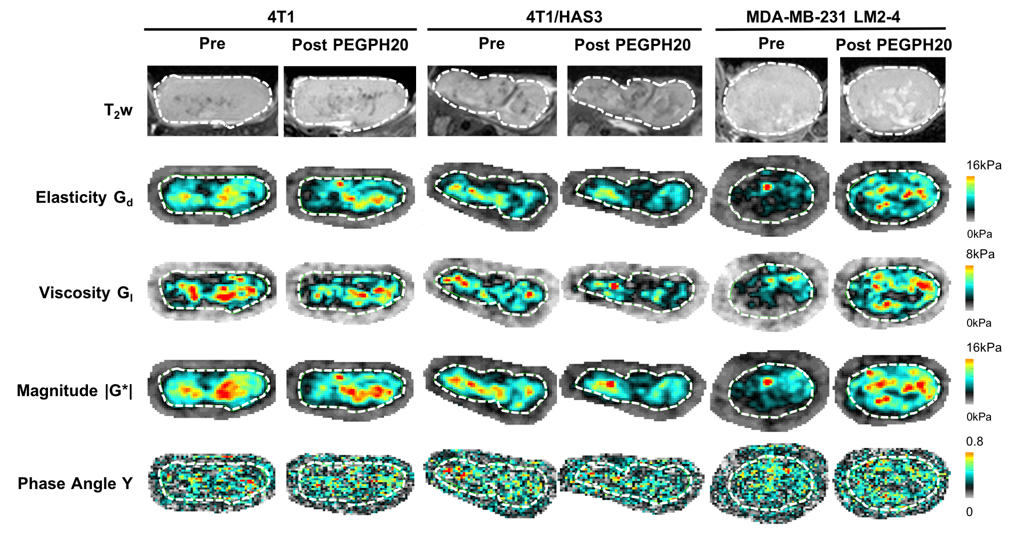
Figure 1. Anatomical T2-weighted (T2w) MRI and parametric maps of Gd, Gl, |G*| and Y for representative 4T1, 4T1/HAS3 and MDA-MB-231 LM2-4 tumours prior to and 24 hours after treatment with PEGPH20 (1 mg/kg). The tumour is delineated by a white dashed line.
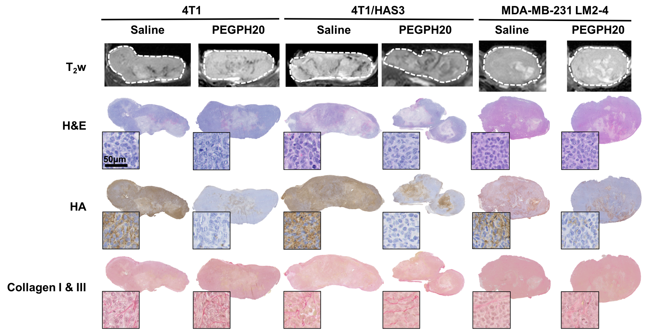
Figure 3. Anatomical T2-weighted (T2w) MRI and aligned tissue sections stained with H&E, HTI-601 (hyaluronan/HA) and picrosirius red (collagen I & III). Representative saline and PEGPH20 treated tumours are shown for each tumour model (4T1, 4T1/HAS3 and MDA-MB-231 LM2-4). The PEGPH20 treated tumours are the same as those shown in Figure 1. The whole section images highlight the close matching of the histology with MRI. Representative high-power images (20x) are inset next to each whole tumour section.