Deepti Upadhyay1, Jinny Sun1, Joao Piraquive Agudelo1, Hongjuan Zhao2, Rosalie Nolley2, Robert Bok1, James D. Brooks2, Donna M. Peehl1, John Kurhanewicz1, and Renuka Sriram1
1Department of Radiology and Biomedical Imaging, University of California, San Francisco, San Francisco, CA, United States, 2Department of Urology, Stanford University, Stanford, CA, United States
1Department of Radiology and Biomedical Imaging, University of California, San Francisco, San Francisco, CA, United States, 2Department of Urology, Stanford University, Stanford, CA, United States
Metabolic characterization of renal cell carcinoma patient derived-xenografts and tissue slice cultures using stable isotope resolved metabolism showed metabolic heterogeneity among PDXs and TSCs of different clinical and pathological stages.
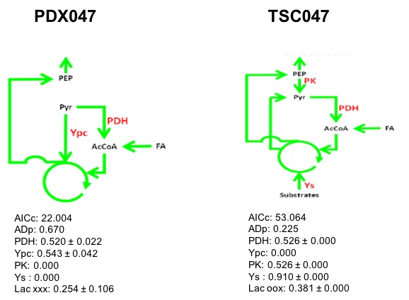
Figure 4. 13C Isotopomer modeling of [U-13C] glucose-labeled of PDX047 and TSC047 using tcaCALC software. (PDH - Pyruvate dehydrogenase, PK-Pyruvate Kinase, Y-denotes anaplerotic denotes, Ypc - Pyruvate Carboxylase and Ys- anaplerosis via succinyl CoA, glutaminolysis)
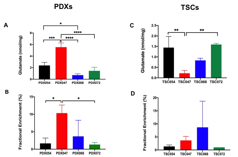
Figure 3: Comparison of (A) steady state concentration and (B) Fractional enrichment of glutamate among PDX model, while (C) & (D) represent steady state concentration and Fractional enrichment of glutamate, respectively among TSCs model. *p<0.05, **p<0.005, ***p< 0.0005, n=3 for all groups except TSC072 (n=2)