Farzad Alizadeh1,2, Anahita Fathi Kazerooni3,4, Hanieh Bahrampour5, Hanieh Mobarak Salari1,2, and Hamidreza Saligheh Rad1,2
1Department of Medical Physics and Biomedical Engineering, Tehran university of Medical Science, Tehran, Iran (Islamic Republic of), 2Quantitative MR Imaging and Spectroscopy Group, Research Center for Molecular and Cellular Imaging, Tehran, Iran (Islamic Republic of), 3Center for Biomedical Image Computing and Analytics (CBICA), University of Pennsylvania, Philadelphia, PA, United States, 4Department of Radiology, Perelman School of Medicine, University of Pennsylvania, Philadelphia, PA, United States, 5Biomaterials Engineering, School of Metallurgy and Materials Engineering, Iran University of Science and Technology, Tehran, Iran (Islamic Republic of)
1Department of Medical Physics and Biomedical Engineering, Tehran university of Medical Science, Tehran, Iran (Islamic Republic of), 2Quantitative MR Imaging and Spectroscopy Group, Research Center for Molecular and Cellular Imaging, Tehran, Iran (Islamic Republic of), 3Center for Biomedical Image Computing and Analytics (CBICA), University of Pennsylvania, Philadelphia, PA, United States, 4Department of Radiology, Perelman School of Medicine, University of Pennsylvania, Philadelphia, PA, United States, 5Biomaterials Engineering, School of Metallurgy and Materials Engineering, Iran University of Science and Technology, Tehran, Iran (Islamic Republic of)
Convolutional neural networks are able to differentiate brain tumorous tissue subregions based on 1H-MRS data with acceptable accuracy scores.
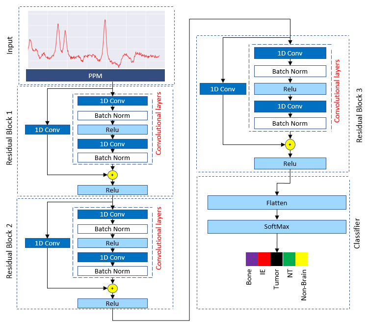
Our proposed Resnet-based convolutional neural
network architecture applied to classify five tissue types in diffuse glioma.

T1-w image of a patient with a diffuse glioma (a).
Chemical Shift Image (CSI) of the patient (b). Rotated overlaid image of CSI
grid on T1-w image (c). color coded grid of CSI in order to exact localization of the signals (d).