Emma Bluemke1, Ambre Bertrand1, Kwun-Ye Chu2,3, Nigar Syed2, Andrew Murchison3,4, Tessa Greenhalgh3,5, Brian Burns6, Martin Craig7, Nia Taylor3, Ketan Shah2,3, Fergus Gleeson2,3, and Daniel Bulte1
1University of Oxford, Oxford, United Kingdom, 2MRC Oxford Institute for Radiation Oncology, Department of Oncology, University of Oxford, Oxford, United Kingdom, 3Radiotherapy Department, Oxford University Hospitals NHS, Oxford, United Kingdom, 4Nuffield Department of Medicine, University of Oxford, Oxford, United Kingdom, 5University Hospital Southampton NHS FT, Southhampton, United Kingdom, 6GE Healthcare, Menlo Park, CA, United States, 7University of Nottingham, Nottingham, United Kingdom
1University of Oxford, Oxford, United Kingdom, 2MRC Oxford Institute for Radiation Oncology, Department of Oncology, University of Oxford, Oxford, United Kingdom, 3Radiotherapy Department, Oxford University Hospitals NHS, Oxford, United Kingdom, 4Nuffield Department of Medicine, University of Oxford, Oxford, United Kingdom, 5University Hospital Southampton NHS FT, Southhampton, United Kingdom, 6GE Healthcare, Menlo Park, CA, United States, 7University of Nottingham, Nottingham, United Kingdom
VFA T1 was higher than the MOLLI T1 by 27%. No significant difference in standard deviation within tumour ROIs from VFA vs MOLLI. VFA was a more robust method, considering patient motion in the z-plane rendered MOLLI unusable for OE-MRI analysis.
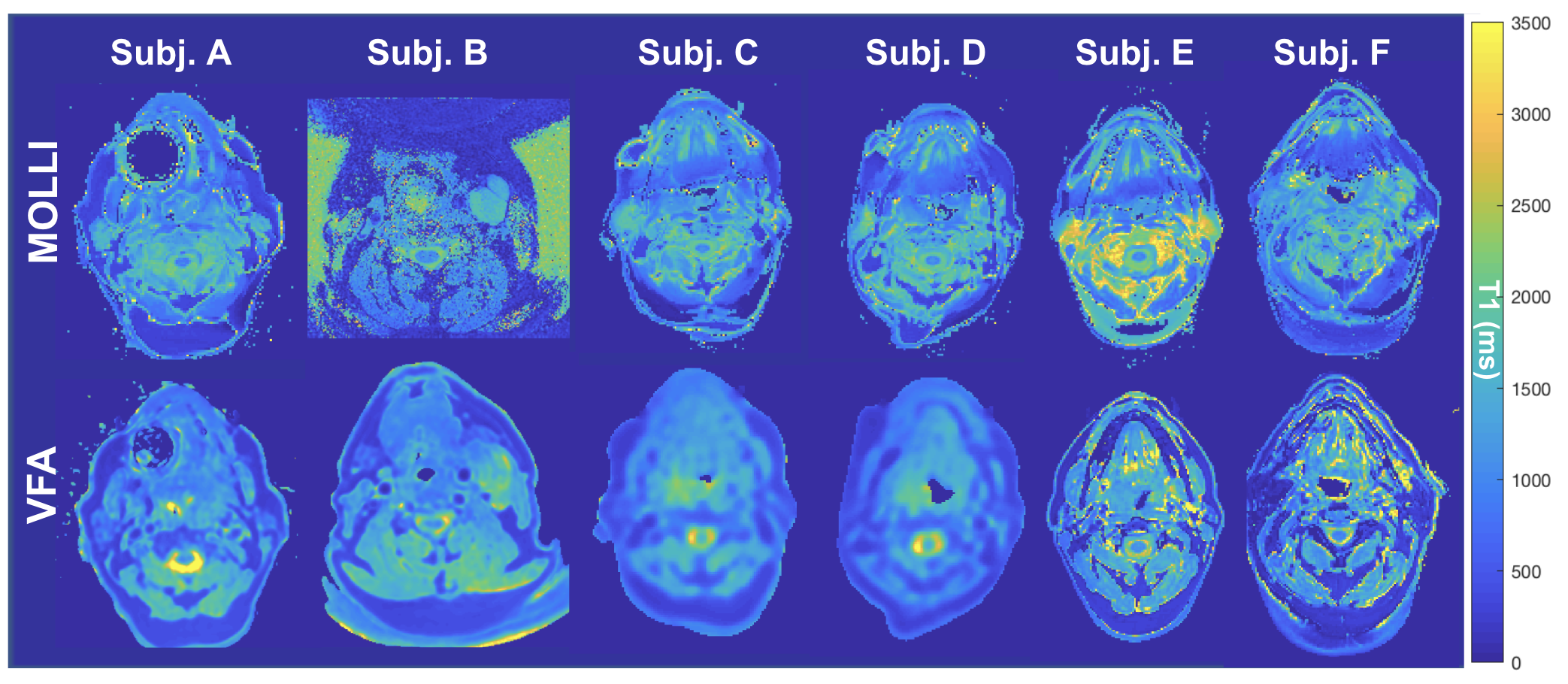
Example VFA and MOLLI T1 maps for 6 subjects (displaying a comparable slice). The field-of-view of the images has been cropped for ease of display.
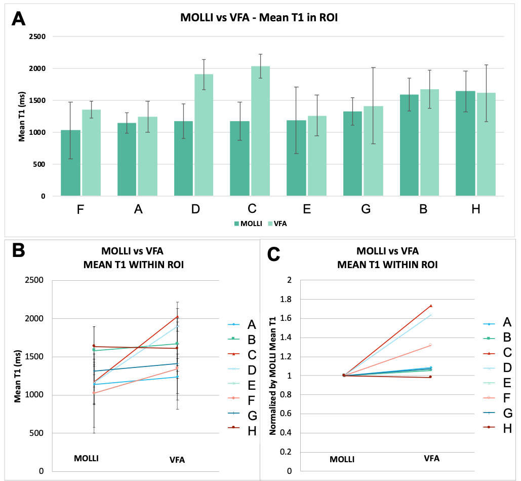
(A-B) The mean T1 in the tumour ROI of all subjects. The error bars represent the standard deviation of T1 values within the tumour ROI. (C) The mean VFA T1, normalized by the respective mean MOLLI T1 for each subject to illustrate the relative change in T1 clearly.