Yifan Yuan1, Qi Yue1, Xiang Zou1, Jiajun Cai1, Ying-Hua Chu2, Yi-Cheng Hsu2, Patrick Alexander Liebig3, Hui Zhang4, He Wang4, Liang Chen1, and Ying Mao1
1Department of Neurosurgery, Huashan Hospital, Fudan University, Shanghai, China, 2MR Collaboration, Siemens Healthcare Ltd., Shanghai, China, 3Siemens Healthcare GmbH, Erlangen, Germany, 4Institute of Science and Technology for Brain-Inspired Intelligence, Fudan University, Shanghai, China
1Department of Neurosurgery, Huashan Hospital, Fudan University, Shanghai, China, 2MR Collaboration, Siemens Healthcare Ltd., Shanghai, China, 3Siemens Healthcare GmbH, Erlangen, Germany, 4Institute of Science and Technology for Brain-Inspired Intelligence, Fudan University, Shanghai, China
We aim to establish a new
grade-discrimination criterion via APT and explore the possible metastatic
margin of diffuse glioma at 7T. With the early exploration of metastatic
margin, the APT imaging at 7T may significantly facilitate the surgical
strategy and surgical excision extension.
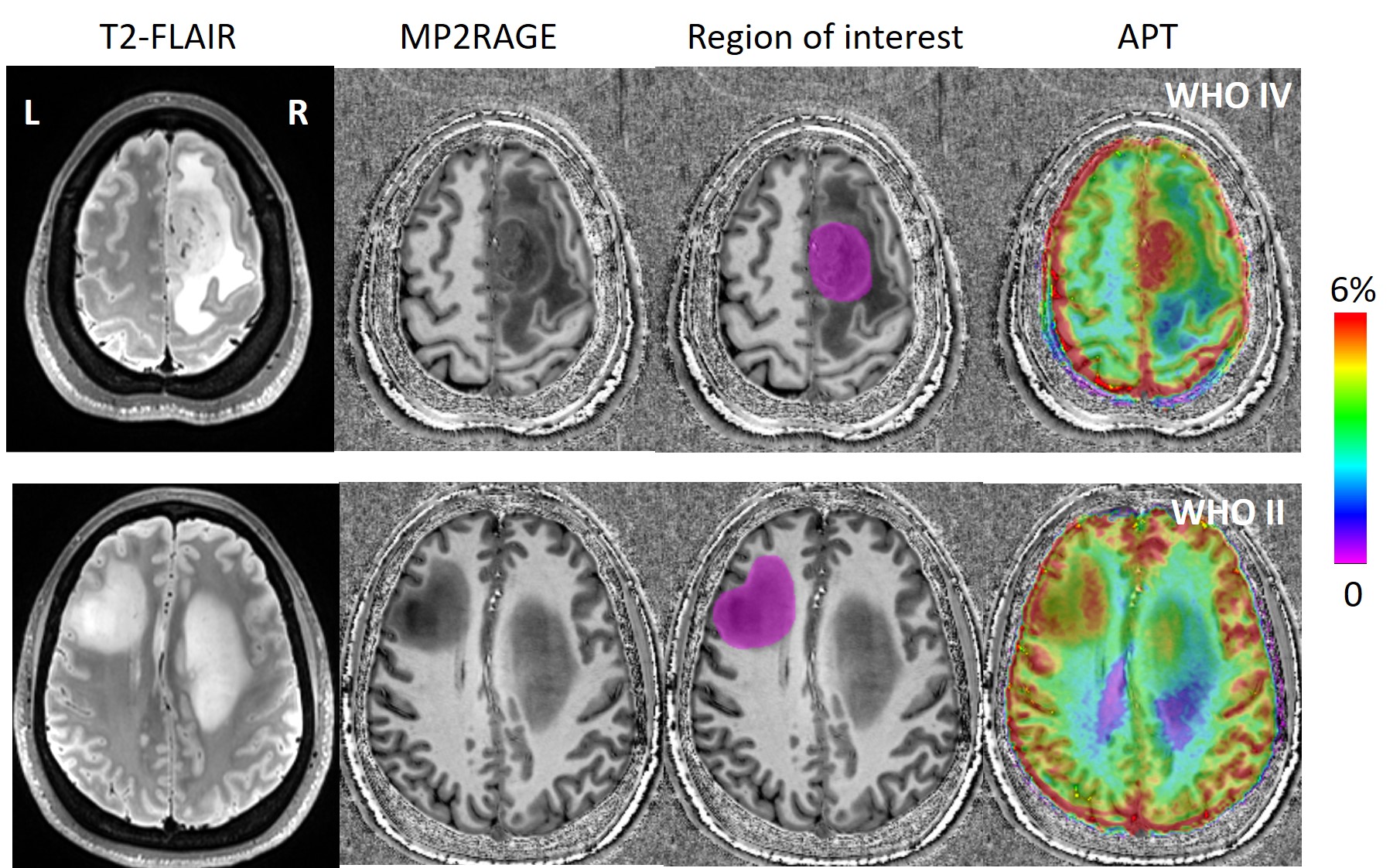
Figure 1.
T2-Flair, MP2RAGE, and ATP were acquired from Subject
9. The images in the top row is from the higher grade (WHO Grade IV) lesion.
The results of the lower grade (WHO Grade II) lesion are shown in the bottom
row. Larger APT values can be found in the high-grade region. Region of
interest (ROI) was defined within the glioma lesion, and the statistical
analysis was performed based on the ROIs.
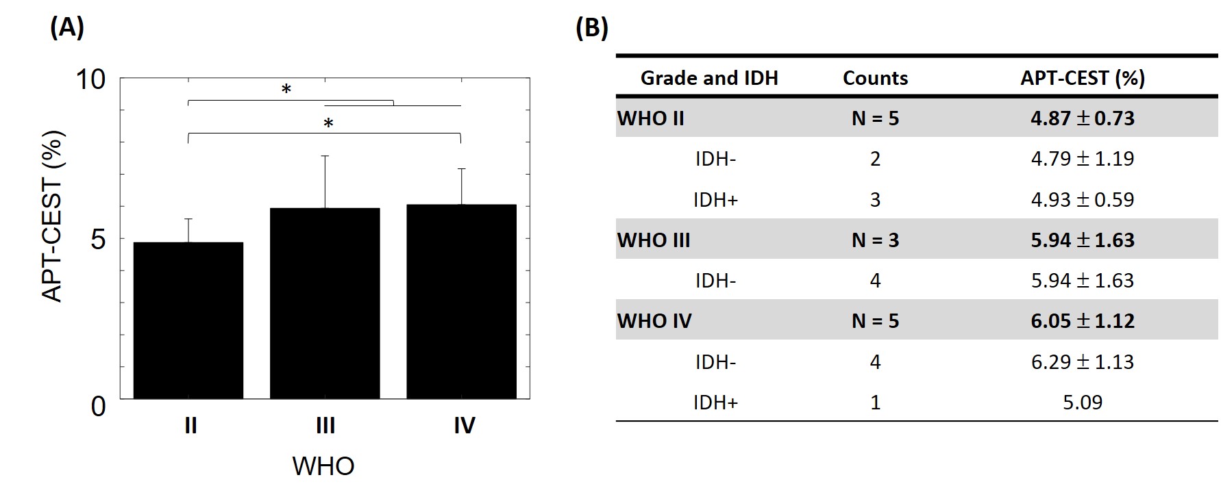
Figure 2.
The group statistic using APT-CEST
signal intensities obtained from all three grades of glioma were shown. (A) The
APT-CEST signal intensity of high-grade (III and IV) glioma is significantly
higher than low-grade (II) glioma (p-value <0.05). There was a significantly
lower APT-CEST signal in WHO grade II compare to WHO grade IV (p-value
<0.05). (B) The IDH mutation status in patient group was classified by WHO.