Anna Morr1, Marcin Nowicki2, Gergely Bertalan1, Rafaela Vieira da Silva1, Carmen Infante Duarte1, Stefan Paul Koch1, Philipp Boehm-Sturm1, Ute Krügel2, Jürgen Braun1, Barbara Steiner1, Josef Käs2, Thomas Fuhs2, and Ingolf Sack1
1Charité - Universitätsmedizin Berlin, Berlin, Germany, 2Universität Leipzig, Leipzig, Germany
1Charité - Universitätsmedizin Berlin, Berlin, Germany, 2Universität Leipzig, Leipzig, Germany
Subregional and microscopic stiffness of the murine dentate
gyrus was detected by in vivo by MRE and ex vivo by atomic force microscopy. Both
methods consistently revealed marked soft-solid properties of the subgranular
zone, a neurogenic niche in mammalians.
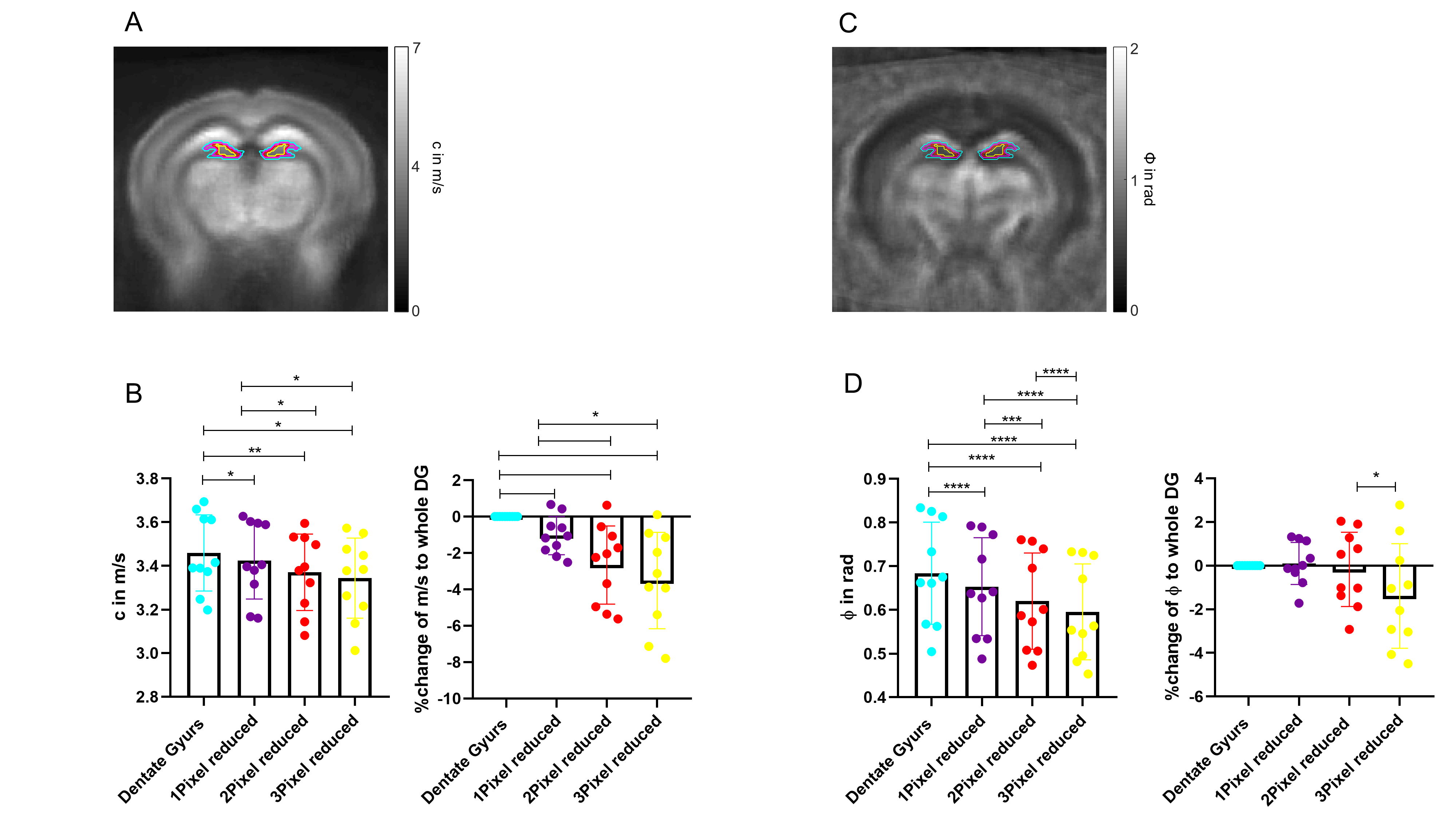
A Representative
c-map showing the DG mask and its diminution in a pixel-wise manner. B there is significant decrease in
stiffness (c in m/s) when down-sizing the mask. C Representative ϕ-map
showing the erosion of the DG mask in a pixel-wise manner. D There is significant decrease in the loss angle, when the mask is
eroded. **** ≦ 0.0001,
*** ≦ 0.001, ** ≤ 0.01,
≤ 0.05*
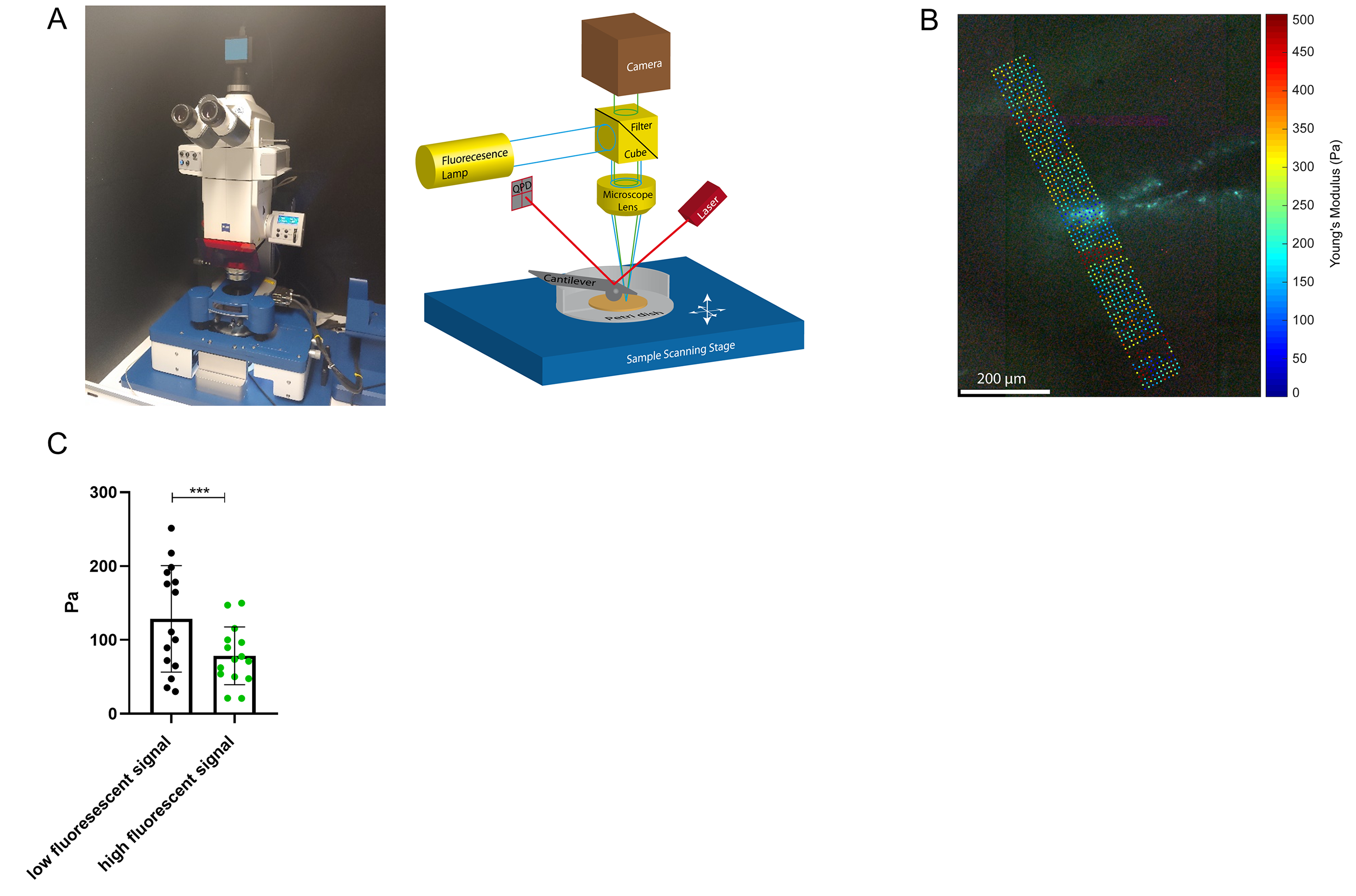
A AFM
set-up on the left, on the right a sketch, the brain slice is placed in
a petri dish on the sample scanner stage of the AFM. The AFM-cantilever is
probing the brain slice from above. An upright fluorescent microscope allows
imaging the GFP-signal, optical and AFM-Image are registered with pixel-precision.
B Exemplary fluorescent image of a brain slice overlaid with the local
Young‘s Modulus. C The SGZ (high fluorescence signal) is softer compared
to neighboring regions(low fluorescence signal) in the dentate gyrus, paired
Wilcoxon-test was performed. *** ≦ 0.001