Fatiha Andoh1, Claire Pellot-Barakat1, and Xavier Maître1
1Université Paris-Saclay, CEA, CNRS, Inserm, BioMaps, Orsay, France
1Université Paris-Saclay, CEA, CNRS, Inserm, BioMaps, Orsay, France
Brain fluid overpressure
and resulting loss of water contents in CSF and orbital compartments were
confirmed by T2 mapping in
head down tilt position. The overall brain mechanical response in such microgravity
analogous conditions, cerebral tissue stiffening, was revealed by whole brain
MRE.
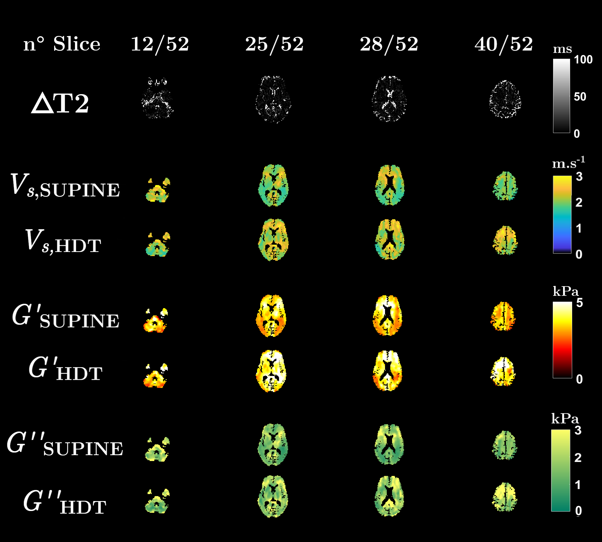
Figure 1: Axial
maps at slices 12, 25, 28 and 40 of the absolute variation of the MR signal lifetime,
ΔT2,
between 0° and 17° positions (top row). ΔT2 is essentially zero everywhere but in the CSF
and orbital compartments where it exhibits a clear T2 decrease
in HDT position. Shear velocity, Vs,
and viscoelastic moduli, G’ and G”, maps reveal a global mechanical increase
between 0° supine and 17° HDT in the cerebral tissues (bottom rows).
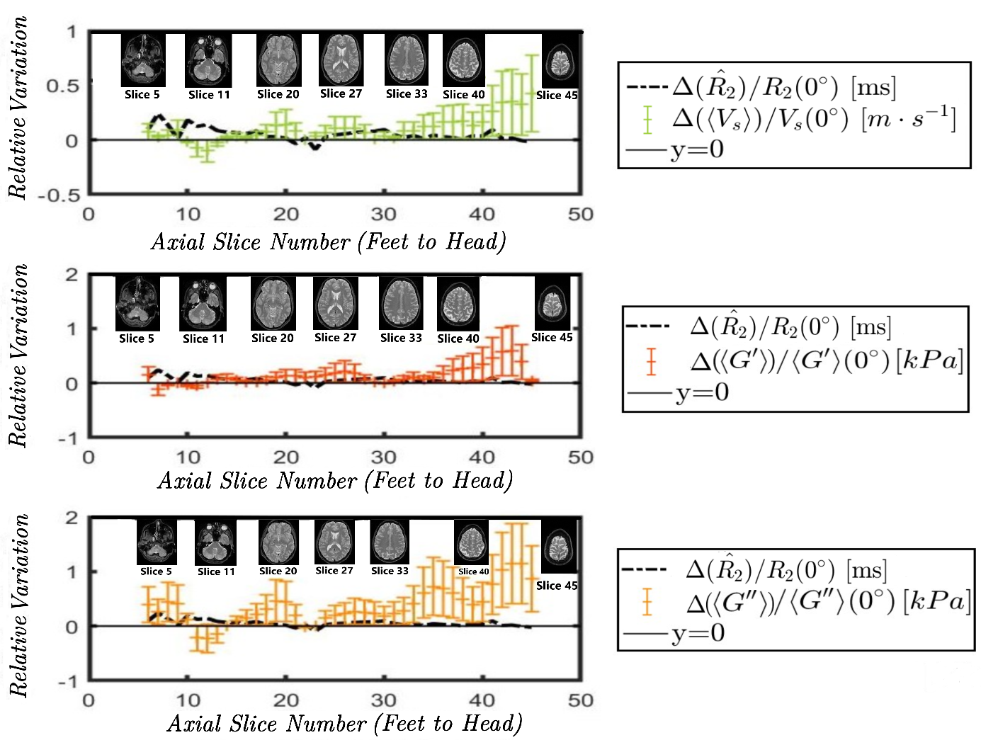
Figure 2: Relative variation of the median
MR transverse relaxivity (R2 = 1/T2) (black), of the mean shear velocity ‹Vs› (green), the mean shear elasticity ‹G’› (dark orange) and shear
viscosity ‹G”› (light orange) across the inferior-superior axial slices. While T2 positively varies only locally in the CSF and
orbital compartments, ‹Vs›, ‹G’› and ‹G”› increase everywhere in the
cerebral tissues, remain rather constant in cerebellum and decrease around the
tentorium.