Dilyana B. Mangarova1,2, Julia Brangsch1, Marcus R. Makowski1,3, Bernd Hamm1, Ingolf Sack1, Jürgen Braun 4, and Gergely Bertalan1
1Department of Radiology, Charité - Universitätsmedizin Berlin, Berlin, Germany, 2Department of Veterinary Pathology, Free University of Berlin, Berlin, Germany, 3Department of Diagnostic and Interventional Radiology, Technical University of Munich, Berlin, Germany, 4Institute for Medical Informatics, Charité - Universitätsmedizin Berlin, Berlin, Germany
1Department of Radiology, Charité - Universitätsmedizin Berlin, Berlin, Germany, 2Department of Veterinary Pathology, Free University of Berlin, Berlin, Germany, 3Department of Diagnostic and Interventional Radiology, Technical University of Munich, Berlin, Germany, 4Institute for Medical Informatics, Charité - Universitätsmedizin Berlin, Berlin, Germany
We demonstrate the feasibility
of MR elastography (MRE) to determine pathologically altered stiffness
in abdominal aortic
aneurysm samples. Aortic wall stiffness is increased and thrombus morphology in
MRE correlates with accumulation of extracellular matrix
components in histology.
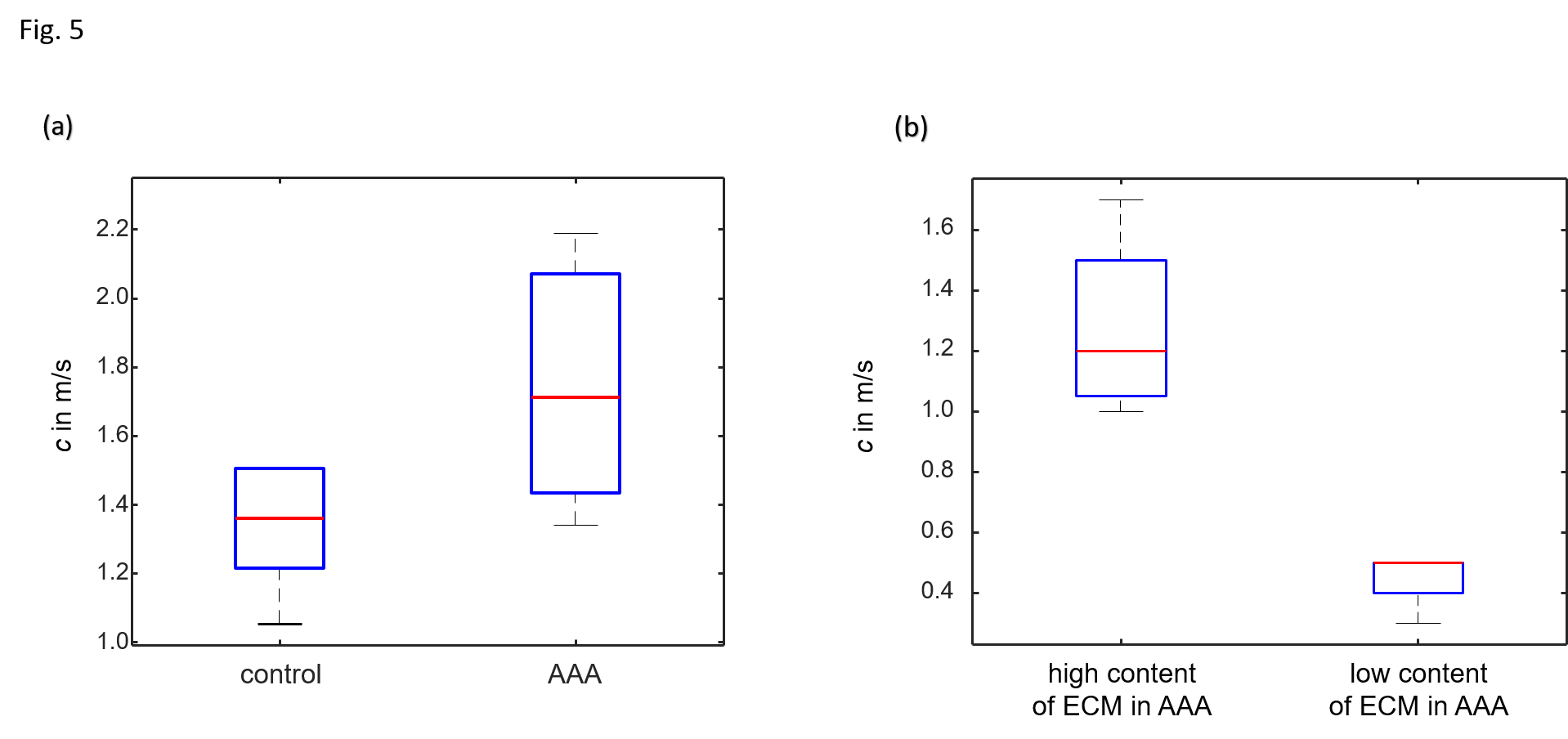
(a) Group values of
SWS of aortic wall of controls and AAA. (b) Regional analysis based on
Elastica-van-Gieson stainings of three AAAs for regions with high (red ROIs)
and low content (blue ROIs) of extracellular matrix (ECM) components.
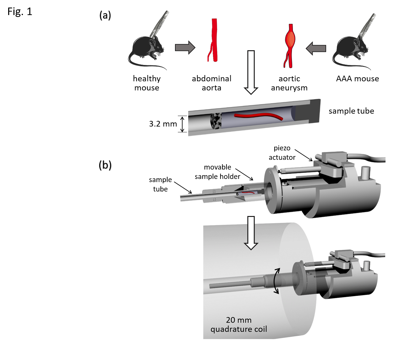
Experimental setup.
(a) Abdominal aorta samples of control mice and mice with AAA were transferred in glass
tubes with an inner diameter of 3.2 mm and embedded in ultrasound gel (details
see text). (b) Upper row: transducer system for vibration generation with
inserted sample tube. Main components of the transducer system include a piezo
actuator which is connected by a transducer rod to a movable sample holder.
Lower row: position of the transducer system in the quadrature volume coil with
inner diameter of 20 mm.