Shruti Mishra1,2, Bin Deng2,3,4, W. Scott Hoge1,2,3, Giacomo Annio5, Ralph Sinkus5, and Samuel Patz1,2
1Department of Radiology, Brigham & Women's Hospital, Boston, MA, United States, 2Harvard Medical School, Boston, MA, United States, 3Athinoula A. Marginos Center for Biomedical Imaging, Charlestown, MA, United States, 4Radiology, Massachusetts General Hospital, Boston, MA, United States, 5Laboratory for Vascular Translational Science (LVTS), Institut National de la Santé et de la Recherche Médicale (INSERM), Paris, France
1Department of Radiology, Brigham & Women's Hospital, Boston, MA, United States, 2Harvard Medical School, Boston, MA, United States, 3Athinoula A. Marginos Center for Biomedical Imaging, Charlestown, MA, United States, 4Radiology, Massachusetts General Hospital, Boston, MA, United States, 5Laboratory for Vascular Translational Science (LVTS), Institut National de la Santé et de la Recherche Médicale (INSERM), Paris, France
High resolution MRE of the brain in healthy volunteer subjects results in a lower mean estimation of wavelength (and therefore stiffness) for a given brain region and greater discriminability between adjacent cortical gray matter and subcortical white matter.
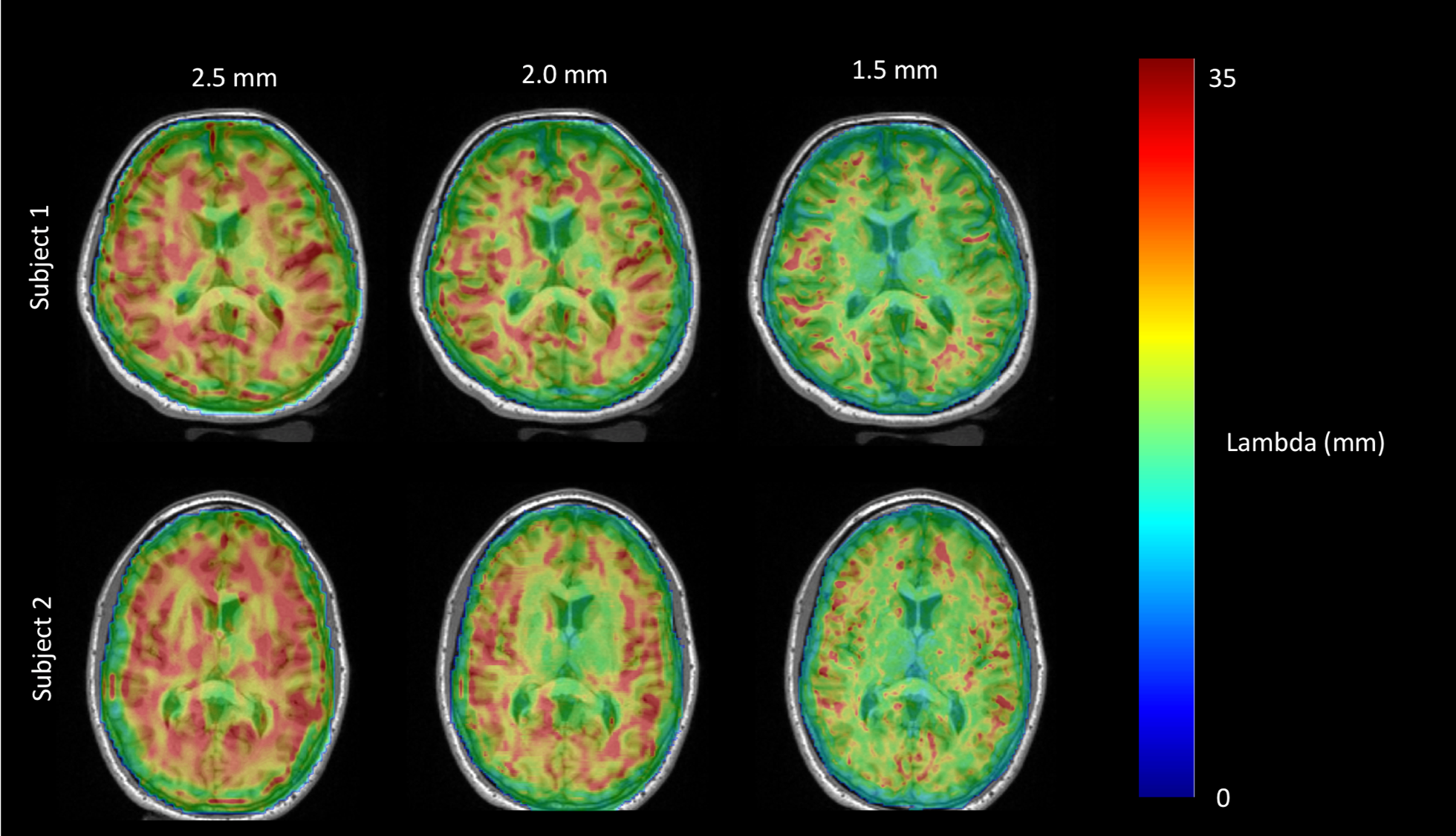
Figure 1: Estimated wavelength ($$$\lambda$$$) overlaid on high-resolution T1 MPRAGE for MRE sequences obtained at 2.5 mm (left), 2.0 mm (middle), and 1.5 mm (right) isotropic resolutions on two healthy adult subjects.
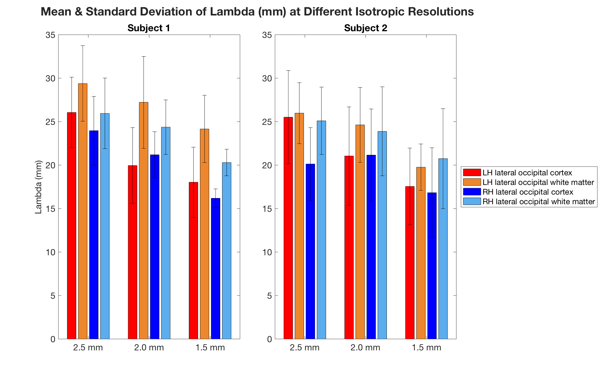
Figure 3: Mean and standard deviation of the estimated wavelength (mm) in each of the left and right hemisphere lateral occipital cortex ROIs and adjacent white matter, depicted in Figure 2, from MRE scans done at 2.5, 2.0, and 1.5 mm resolutions, respectively, for each of the two subjects.