Jiahui Li1, Prachi Singh2, Marzanna Obrzut1, Xin Lu1, Kevin J. Glaser1, Alina Allen3, Sudhakar K. Venkatesh1, Taofic Mounajjed4, Jun Chen1, Armando Manduca1, Vijay Shah3, Richard L. Ehman1, and Meng Yin1
1Radiology, Mayo Clinic, Rochester, MN, United States, 2Sleep and Cardiometabolic Health, Pennington Biomedical Research Center, Baton Rouge, LA, United States, 3Gastroenterology, Mayo Clinic, Rochester, MN, United States, 4Anatomic Pathology, Mayo Clinic, Rochester, MN, United States
1Radiology, Mayo Clinic, Rochester, MN, United States, 2Sleep and Cardiometabolic Health, Pennington Biomedical Research Center, Baton Rouge, LA, United States, 3Gastroenterology, Mayo Clinic, Rochester, MN, United States, 4Anatomic Pathology, Mayo Clinic, Rochester, MN, United States
MRE-based assessment of subcutaneous adipose tissue
provides biomarkers that show promise for improving the diagnosis of NASH and
assessing systemic metabolic dysfunction in obese patients.
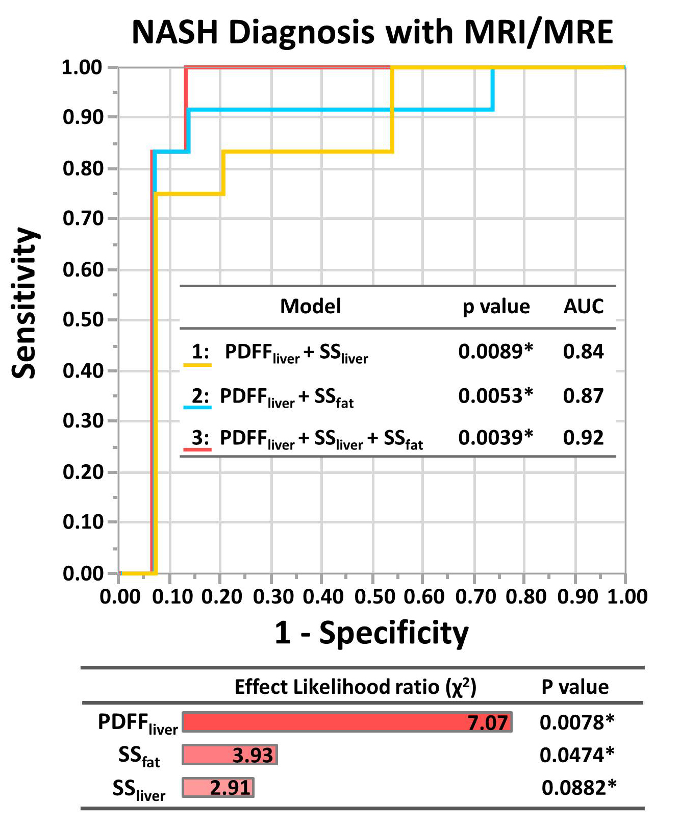
Figure 3. Receiver
Operating Characteristic (ROC) of NASH prediction models and effect test for various
models. ROC analyses for the
diagnosis of NASH based on the nominal logistic fit models. PDFFliver, fat fraction of liver; SSliver, liver stiffness; SSfat, subcutaneous fat stiffness. *, p < 0.05.
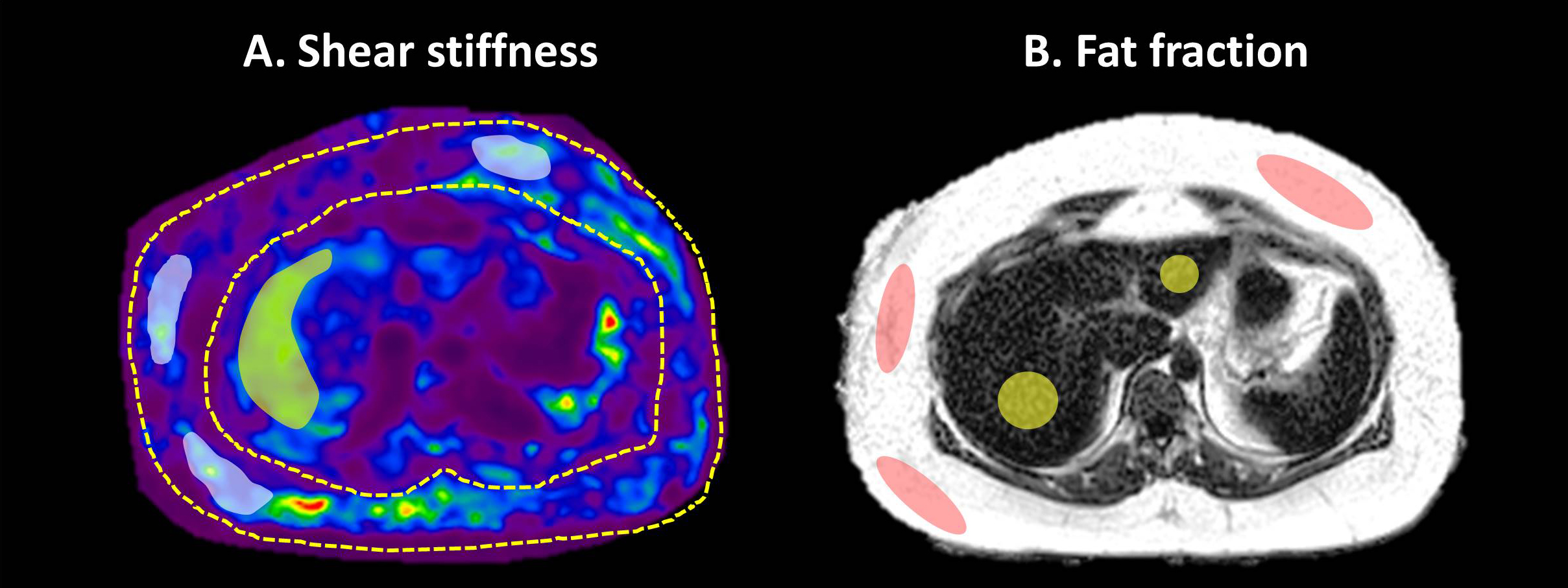
Figure 1. Examples of
ROI placement to measure liver and fat shear stiffness and fat fraction. The
white area (A) and pink area (B) represent the ROIs used to report the shear
stiffness and fat fraction, respectively, for subcutaneous adipose tissue. The
yellow areas (A and B) represent the ROIs for the liver.