Yuan Le1, Jun Chen1, Phillip J. Rossman1, Ziying Yin1, Kevin J. Glaser1, Joshua D. Trzasko1, Yi Sui1, Stephan Kannengiesser2, Bradley D. Bolster, Jr.3, Joel P. Felmlee1, and Richard L. Ehman1
1Radiology, Mayo Clinic, Rochester, MN, United States, 2Siemens Healthcare GmbH, Erlangen, Germany, 3Siemens Medical Solutions USA, Inc., Salt Lake City, UT, United States
1Radiology, Mayo Clinic, Rochester, MN, United States, 2Siemens Healthcare GmbH, Erlangen, Germany, 3Siemens Medical Solutions USA, Inc., Salt Lake City, UT, United States
This study assessed the feasibility of visualizing shear
strain in intervertebral discs using MR elastography methods, towards a goal of
using this information as a way to quantitatively assess disc degeneration.
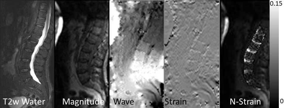
Figure 2. (Left to right) Anatomical T2w water image, MRE
magnitude image, MRE phase difference image, strain images and N-strain map.
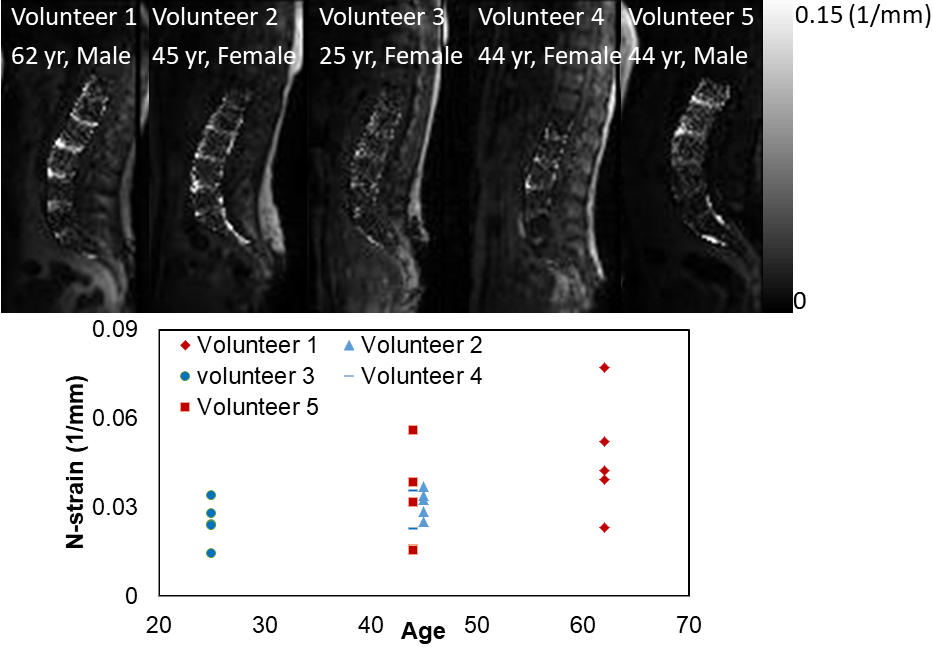
Figure 4 N-strain maps and the N-strain values vs. volunteer’s age.