Abrar Faiyaz1, Irteza Enan Kabir1, Marvin M Doyley1, Ingolf Sack2, Md Nasir Uddin3, and Giovanni Schifitto3
1Department of ECE, University of Rochester, Rochester, NY, United States, 2Department of Radiology, Charité - Universitätsmedizin Berlin, Berlin, Germany, 3Department of Neurology, University of Rochester, Rochester, NY, United States
1Department of ECE, University of Rochester, Rochester, NY, United States, 2Department of Radiology, Charité - Universitätsmedizin Berlin, Berlin, Germany, 3Department of Neurology, University of Rochester, Rochester, NY, United States
MR-Elastography has the potential to detect HIV related brain injury by abnormally low stiffness values. The study explored this potential and reports promising results compared to the diffusion derived metrics with DTI or NODDI.
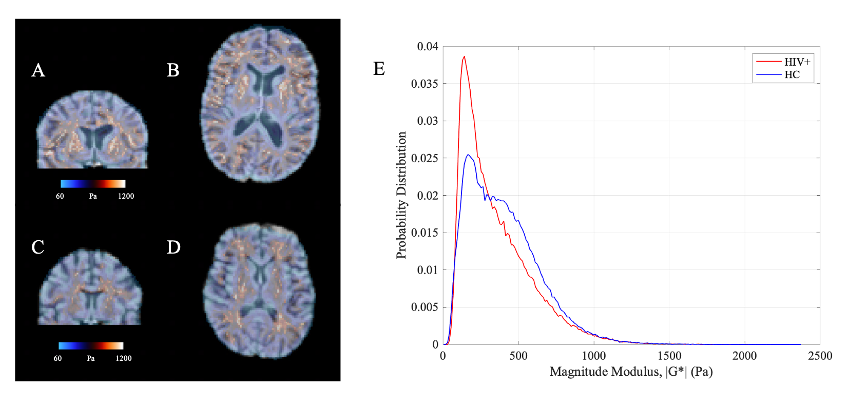
Figure 1: Representative stiffness |G*| maps (both coronal and axial views) from a healthy control (A, B) and HIV+ subject (C, D) with respective probability histograms (E) overlayed on respective T1w images. Intensity scales are also shown. HC: healthy control; HIV+: HIV-infected individual.
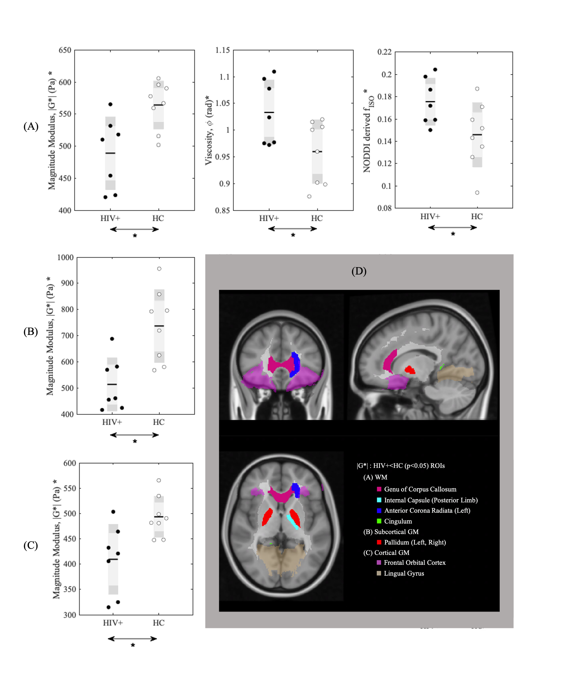
Figure 4: Significant |G*|, and fISO changes in GM and WM ROIs: The group comparison for significant regions of (A) WM, (B) Subcortical GM and (C) Cortical GM are shown. Significant regions for A, B and C are overlayed with respective legends in (D) on standard space with Coronal, Sagittal and Axial view. Relevant parameters are |G*|, $$$\phi$$$ and fISO, only significant changes are shown.