Helge Herthum1, Hugo Carrillo2, Axel Osses2, Sergio Uribe3, Ingolf Sack1, and Cristóbal Bertoglio4
1Experimentelle Radiologie, Charité Universitätsmedizin Berlin, Berlin, Germany, 2Center for Mathematical Modeling, Universidad de Chile, Santiago, Chile, 3Department of Radiology, School of Medicine, Pontificia Universidad Católica de Chile, Santiago, Chile, 4Bernoulli Institute, University of Groningen, Groningen, Netherlands
1Experimentelle Radiologie, Charité Universitätsmedizin Berlin, Berlin, Germany, 2Center for Mathematical Modeling, Universidad de Chile, Santiago, Chile, 3Department of Radiology, School of Medicine, Pontificia Universidad Católica de Chile, Santiago, Chile, 4Bernoulli Institute, University of Groningen, Groningen, Netherlands
Applying the OMME method to MRE in phantom and in vivo experiments provides
wrap-free phase-contrast images with high motion-to-noise ratio. Reconstructed
shear wave speed maps show better detail resolution by overcoming the
unwrapping problem in MRE.
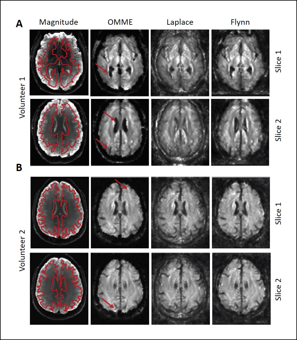
SWS maps for OMME (MEGs=4,8,16,32mT/m) and
Laplace and Flynn unwrapping (MEG=32mT/m) at different slices. The anatomical
reference from MRE magnitude is included (region of interest demarcated in red
for noise estimation). Red arrows indicate where OMME shows greater contrast in
the SWS map. A) Volunteer 1 for
vibration frequency 25Hz at two different slices. Grayscale ranges from 0.3-1.8m/s. B)
Volunteer 2 for vibration frequency 30Hz at two different slices. Grayscale ranges
from 0.2-2.2 m/s.
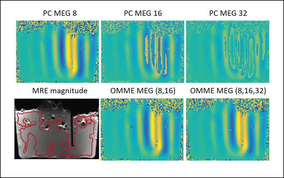
Multiple motion encoding reconstruction in the
phantom for two MEGs 8 and 16 mT/m and three MEGs 8, 16 and 32 mT/m in
comparison to phase contrast images. Color scale is set in the interval 80% of the
dynamic range of the MEG=8 image. Large positive values are colored in yellow
and negative values in blue, while green being zero. The bottom left image
shows the T2 weighted MRE magnitude as geometric reference.