Suhao Qiu1, Zhao He1, Runke Wang1, Ruokun Li2, Fuhua Yan2, and Yuan Feng1
1the Institute for Medical Imaging Technology, Shanghai Jiao Tong University, Shanghai, China, 2the Department of Radiology, Ruijin Hospital, Shanghai, China
1the Institute for Medical Imaging Technology, Shanghai Jiao Tong University, Shanghai, China, 2the Department of Radiology, Ruijin Hospital, Shanghai, China
An electromagnetic
actuator was designed, tested, and verified for brain magnetic resonance
elastography (MRE). The actuator is easy to use, comfort to wear, and can carry
out multi-frequency MRE with high accuracy.
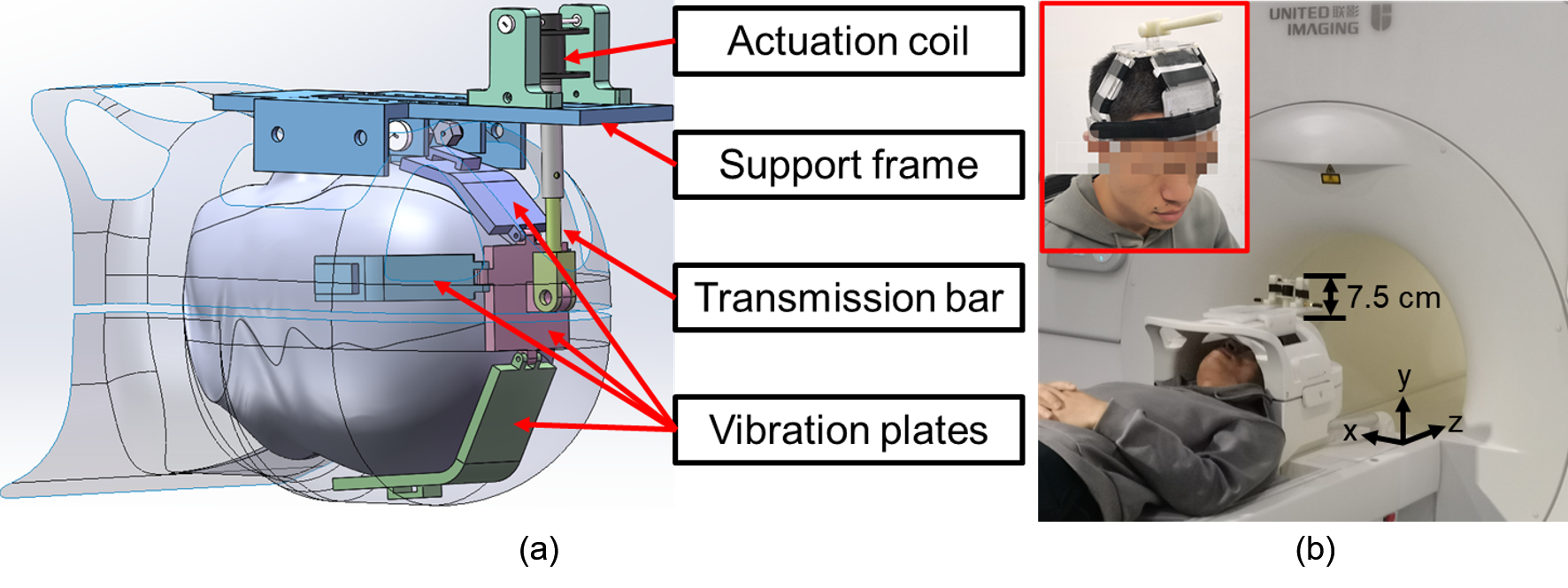
Figure
1. (a)
A 3D rendering of the electromagnetic actuator for brain MRE in working
position. The vibration plates wrap around the human head inside a head coil.
(b) A volunteer wearing the actuator before moving into the MR bore for MRE.
The vibration plates are fixed on the head with soft bandages. The part of the
actuator that extrudes out of the head coil is 7.5 cm in height, leaving enough
space for positioning.
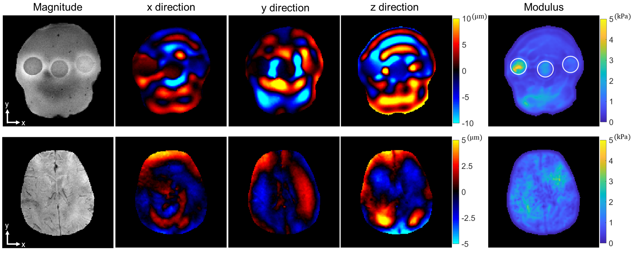
Figure 5. The
first line and the second line are the typical MRE results from the phantom and a volunteer, respectively. Column 1 is the magnitude
image. Column 2-4 are the real part of the first harmonic component of the
displacement field in x, y, and z direction, respectively. Column 5 is the LFE inversion results after smoothing. The locations of these three
agar inclusions are indicated by white circles.