Helge Herthum1, Heiko Tzschätzsch1, Tom Meyer1, Mehrgan Shahryari1, Lisa Stencel1, Jing Guo1, Jürgen Braun2, and Ingolf Sack1
1Experimentelle Radiologie, Charité Universitätsmedizin Berlin, Berlin, Germany, 2Institut für medizinische Informatik, Charité Universitätsmedizin Berlin, Berlin, Germany
1Experimentelle Radiologie, Charité Universitätsmedizin Berlin, Berlin, Germany, 2Institut für medizinische Informatik, Charité Universitätsmedizin Berlin, Berlin, Germany
We here present multifrequency wavenumber analysis for
MR elastography of the human brain in 2D and 3D. Reproducibility and detail
resolution of the new methods are better than obtained from standard multifrequency
inversion methods.
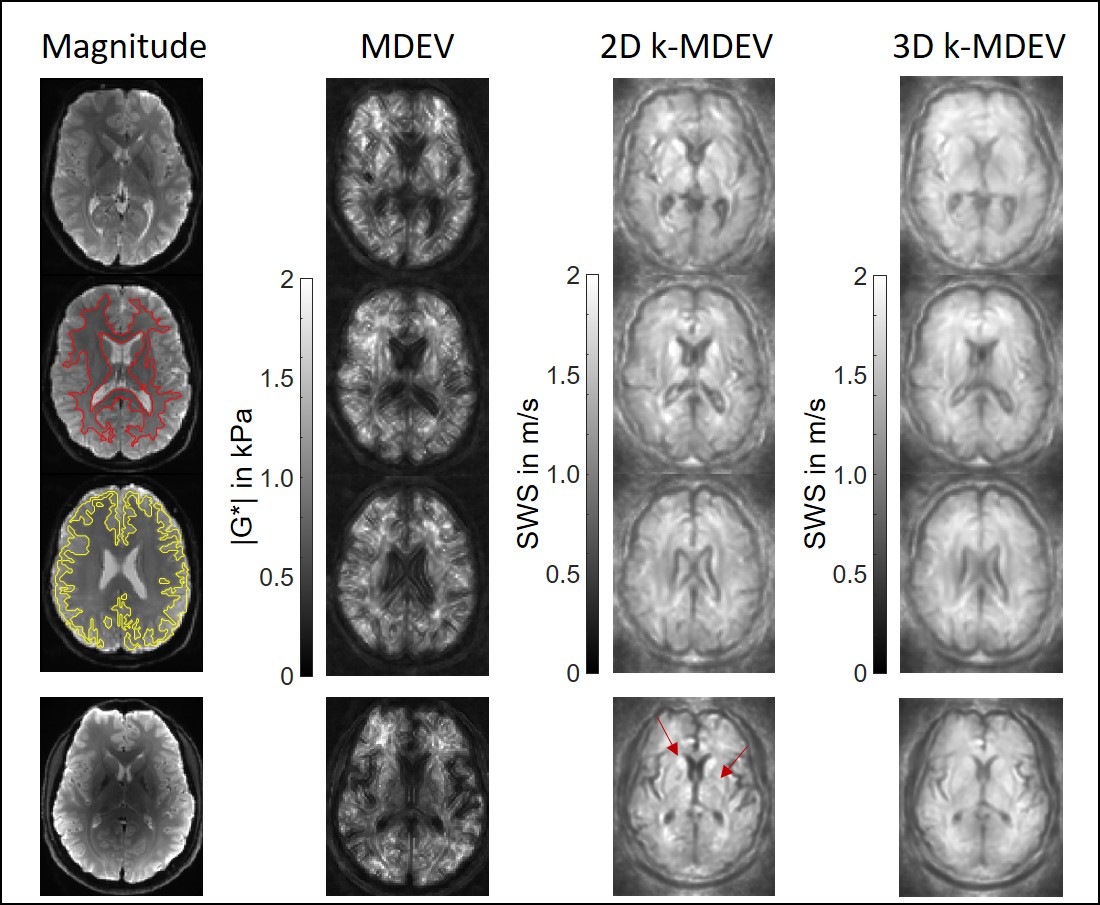
Results from MDEV (|G*|) and 2D and 3D k-MDEV (SWS) inversion in different
slices (1.5-Tesla). Magnitude images are shown on the left side together with
segmented regions for white matter (red) and gray matter (yellow). The last row
shows results for the measurement at 3.0-Tesla with 1.6x1.6x2mm3 voxel
size. Increased resolution allows to easily identify anatomical regions in deep gray matter characterized by stiff
properties such as the putamen and caudate nucleus (indicated by arrows).
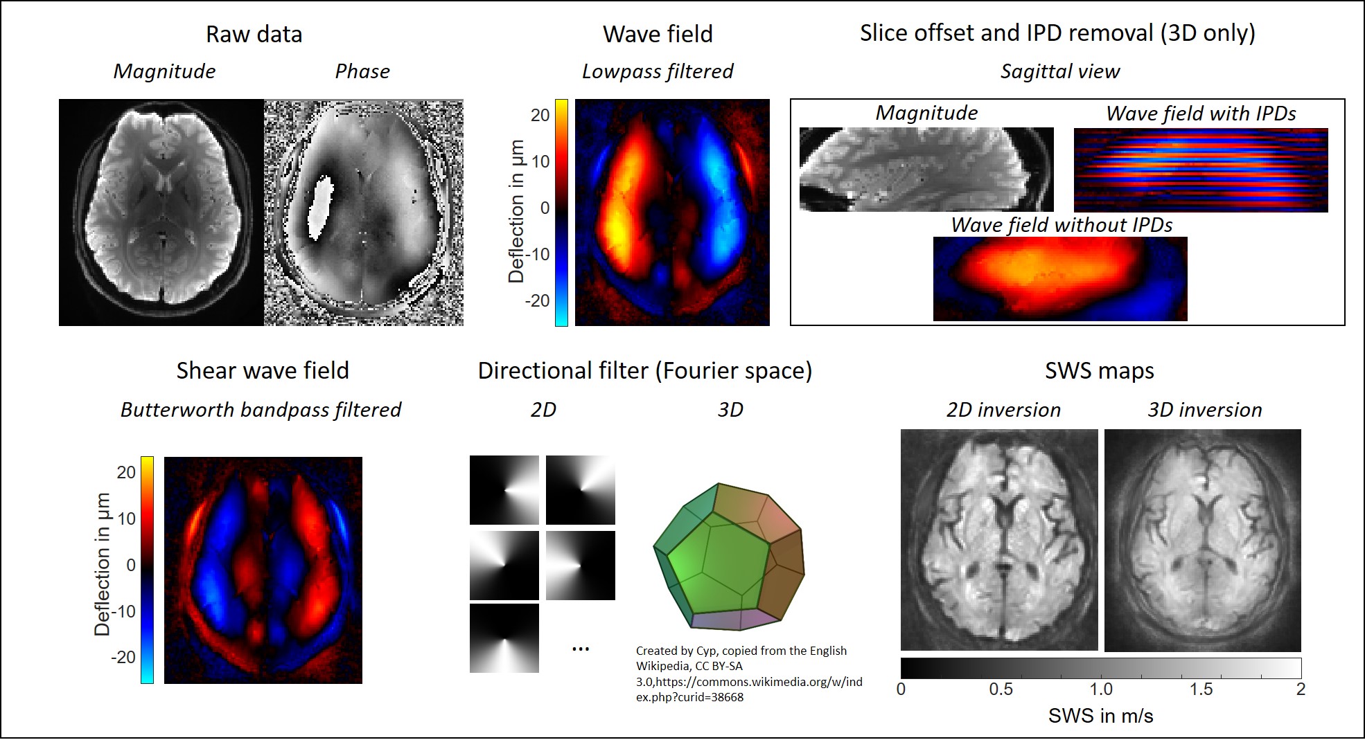
2D and 3D k-MDEV. The phase of the MRE data is smoothed
(Butterworth lowpass, threshold 250 m-1, order 3) and the harmonic
wave field is extracted using the Fourier transform. For 3D, inter-phase
discontinuities (IPD) and slice offsets [13] are removed as shown in the sagittal view. A bandpass
filter (Butterworth, lower threshold 15 m-1, upper threshold 200 m-1,
order 3) suppresses compression waves and the remaining shear wave field is
directionally filtered in 8 (2D) or 20 (3D, right, dodecahedron) directions.
SWS is reconstructed from the phase gradient in 2D and 3D.