Ledia Lilaj1, Helge Herthum2, Tom Meyer2, Mehrgan Shahryari2, Gergely Bertalan2, Alfonso Caiazzo2, Jürgen Braun3, Thomas Fischer2, Sebastian Hirsch4,5, and Ingolf Sack2
1Radiology, Charité – Universitätsmedizin Berlin, Berlin, Germany, 2Charité – Universitätsmedizin Berlin, Berlin, Germany, 3Institute of Medical Informatics, Charité – Universitätsmedizin Berlin, Berlin, Germany, 4Berlin Center for Advanced Neuroimaging, Charité – Universitätsmedizin Berlin, Berlin, Germany, 5Bernstein Center for Computational Neuroscience, Berlin, Germany
1Radiology, Charité – Universitätsmedizin Berlin, Berlin, Germany, 2Charité – Universitätsmedizin Berlin, Berlin, Germany, 3Institute of Medical Informatics, Charité – Universitätsmedizin Berlin, Berlin, Germany, 4Berlin Center for Advanced Neuroimaging, Charité – Universitätsmedizin Berlin, Berlin, Germany, 5Bernstein Center for Computational Neuroscience, Berlin, Germany
Inversion recovery
magnetic resonance elastography (IR-MRE) performs MRE while nulling a tissue
signal based on its T1. In the brain, it reduced the effect of fluid vibration
in CSF-filled areas and improved stiffness estimation in the cortex.
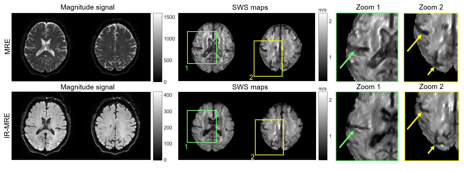
Representative brain images obtained by MRE and IR-MRE in the same
volunteer. The magnitude signals in IR-MRE show solid tissue without freely moving
water, leading to significantly improved depiction of fluid-solid boundaries in
SWS maps (arrows). Note that sulci and ventricles are narrower in IR-MRE than
MRE due to suppressed phase discontinuities, similar to the phantom experiments
shown in Figure 3.
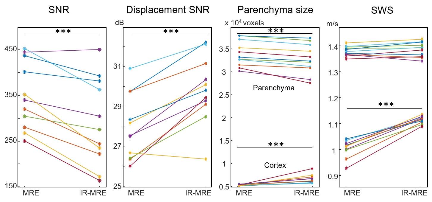
Group values of parameters quantified in this study. *** p < 0.001.