Bin Deng1,2,3, W. Scott Hoge3,4, Shruti Mishra3,4, Giacomo Annio5, Ralph Sinkus5, and Samuel Patz3,4
1Athinoula A. Martinos Center for Biomedical Imaging, Massachusetts General Hospital, Charlestown, MA, United States, 2Radiology, Massachusetts General Hospital, Boston, MA, United States, 3Harvard Medical School, Boston, MA, United States, 4Radiology, Brigham and Women’s Hospital, Boston, MA, United States, 55. Laboratory for Vascular Translational Science (LVTS, Institut National de la Santé et de la Recherche Médicale (INSERM), Paris, France
1Athinoula A. Martinos Center for Biomedical Imaging, Massachusetts General Hospital, Charlestown, MA, United States, 2Radiology, Massachusetts General Hospital, Boston, MA, United States, 3Harvard Medical School, Boston, MA, United States, 4Radiology, Brigham and Women’s Hospital, Boston, MA, United States, 55. Laboratory for Vascular Translational Science (LVTS, Institut National de la Santé et de la Recherche Médicale (INSERM), Paris, France
fMRE brain data from a healthy volunteer acquired at 1Hz switching frequency between two interleaved control paradigms at 1.5-mm isotropic high spatial resolution demonstrated both excellent intra- and inter-scan reproducibility of elastography measurements.
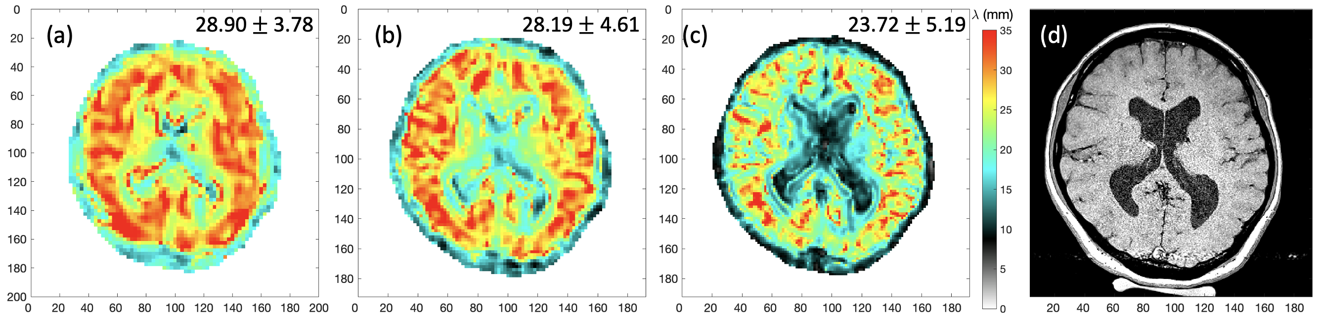
Fig.2: Comparison of reconstructed wavelength maps (λ in mm) of a healthy volunteer acquired at three different spatial resolutions: (a) 2.5 mm isotropic; (b) 2 mm isotropic; and (c) 1.5 mm isotropic voxels. Axial T1 image of 0.5 mm in-plane resolution of the matching slice is shown in (d). Median and standard deviation of valid brain region voxels in the slice are shown on top right corner.
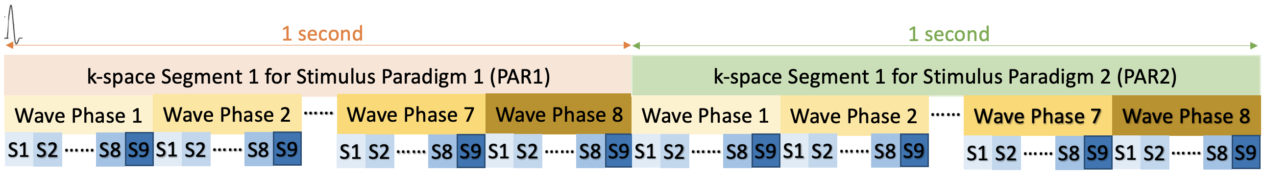
Fig.1: Schematic of fMRE data acquisition with interleaved stimulus paradigms, i.e., PAR1 & PAR2, each containing 9 slices and 8 wave phases. A trigger signal is sent out at the beginning of each block shown to synchronize the k-space acquisition with mechanical vibrations for both paradigms with the loop hierarchy. This block is repeated for each k-space line acquisition and MEG-encoding direction. For fMRE scans with functional activation, a stimulus will be switched ON and OFF in sync with PAR1 and PAR2 sequence timing. In this study, no stimulus is used for either PAR1 or PAR2.