Yang Zhang1,2, Yan-Lin Liu2, Ke Nie1, Jiejie Zhou3, Siwa Chan4, Vivian Youngjean Park5, Min Jung Kim5, Zhongwei Chen3, Jeon-Hor Chen2,4, Meihao Wang3, and Min-Ying Su2
1Department of Radiation Oncology, Rutgers-Cancer Institute of New Jersey, Robert Wood Johnson Medical School, New Brunswick, NJ, United States, 2Department of Radiological Sciences, University of California, Irvine, CA, United States, 3Department of Radiology, The First Affiliate Hospital of Wenzhou Medical University, Wenzhou, China, 4Department of Medical Imaging, Taichung Tzu-Chi Hospital, Taichung, Taiwan, 5Department of Radiology and Research Institute of Radiological Science, Severance Hospital, Yonsei University College of Medicine, Seoul, Korea, Republic of
1Department of Radiation Oncology, Rutgers-Cancer Institute of New Jersey, Robert Wood Johnson Medical School, New Brunswick, NJ, United States, 2Department of Radiological Sciences, University of California, Irvine, CA, United States, 3Department of Radiology, The First Affiliate Hospital of Wenzhou Medical University, Wenzhou, China, 4Department of Medical Imaging, Taichung Tzu-Chi Hospital, Taichung, Taiwan, 5Department of Radiology and Research Institute of Radiological Science, Severance Hospital, Yonsei University College of Medicine, Seoul, Korea, Republic of
These
research shows the potential of Mask R-CNN detection followed by ResNet50
classification for automatic detection and further characterization of
identified lesions to develop a fully-automatic computer-aided diagnosis system
for breast MRI.
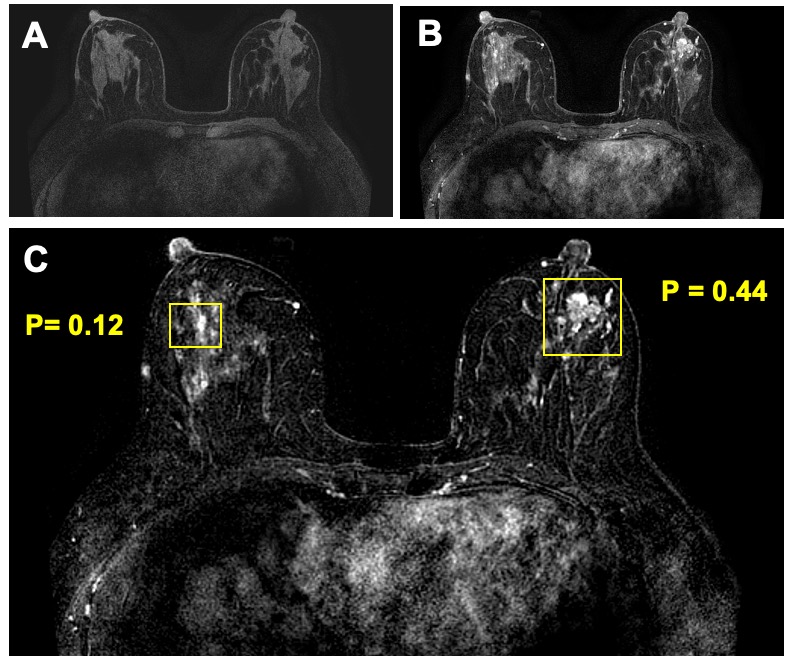
Figure
5: True
negative (TN) case example from a 44-year-old patient with a confirmed benign adenosis in the left breast. Extensive parenchymal enhancements are seen in both breasts. (a) Pre-contrast image acquired using fat-sat sequence; (b) Post-contrast image; (c) Tumor detection results searched by the Mask R-CNN algorithm. Two boxes are generated to identify two suspicious lesions, one in each breast. After evaluation by ResNet50 network, the left lesion has a malignant probability of 0.44, thus correctly diagnosed as benign. The parenchymal enhancements from normal tissues in the right breast has a very low malignant probability of 0.12. These results illustrate the potential of Mask R-CNN detection followed by ResNet50 classification for automatic detection and further characterization of identified lesions to develop a fully-automatic computer-aided diagnosis system for breast MRI.
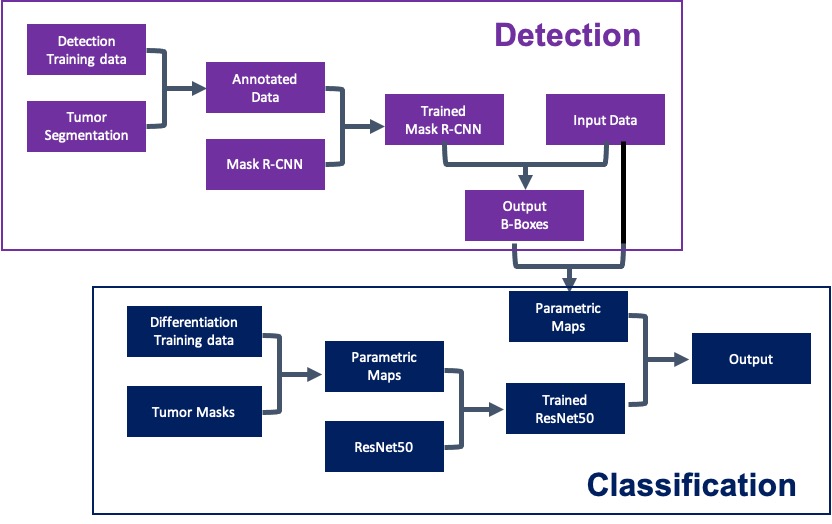
Figure 1: Flow
diagram of the training and testing
courses using Mask R-CNN for
detection (shown in purple) and
ResNet50 for classification (shown in blue).