Lauren K Fang1, Ana E Rodríguez-Soto1, Kathryn E Keenan2, and Rebecca A Rakow-Penner1
1Radiology, University of California San Diego, La Jolla, CA, United States, 2National Institute of Standards and Technology, Boulder, CO, United States
1Radiology, University of California San Diego, La Jolla, CA, United States, 2National Institute of Standards and Technology, Boulder, CO, United States
Overall accuracy and precision of DWI estimates was >88% and improved when normalized by an internal reference. High inter- and intra-scanner variability highlight the need for investigating spatial effects on breast tumor ADC heterogeneity.
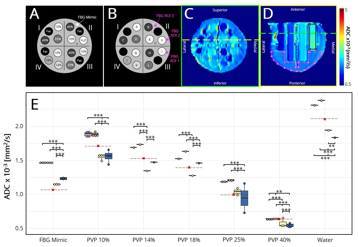
Figure 1. (A) Diffusion breast phantom schematic. Percentages indicate % w/w PVP in water. (B) Schematic with tubes and fibroglandular ROI locations. (C) Axial view of ADC map. Yellow line shows the slice at which ROIs of water (black) and fibroglandular tissue (pink) were drawn in the (D) coronal view. Green line shows the slice at which (C) was taken. (E) Median ADC, shown as dots for each tube. Red lines indicate expected values.6,8
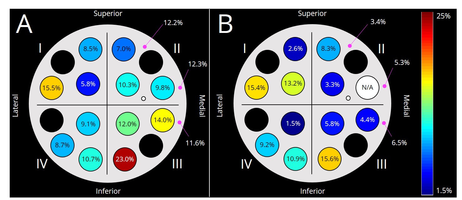
Figure 2. Coefficient of variation across scanners of (A) absolute ADC, and (B) ADC relative to water for each tube.