Anabel M Scaranelo1, Hadassa Degani2, Dov Grobgeld3, Vivianne Freitas1, Shelley Westergard4, Christine Elser5, and Edna Furman-Haran3
1Medical Imaging, University of Toronto, Toronto, ON, Canada, 2DDE MRI Solutions Ltd., Tel Aviv, Israel, 3Weizmann Institute of Science, Rehovot, Israel, 4Princess Margaret Cancer Center, Toronto, ON, Canada, 5University of Toronto, Toronto, ON, Canada
1Medical Imaging, University of Toronto, Toronto, ON, Canada, 2DDE MRI Solutions Ltd., Tel Aviv, Israel, 3Weizmann Institute of Science, Rehovot, Israel, 4Princess Margaret Cancer Center, Toronto, ON, Canada, 5University of Toronto, Toronto, ON, Canada
A prospective feasibility study of breast DTI in high-risk lactating patients indicated high diagnostic sensitivity and specificity and a more accurate cancer detection rate than breast DCE imaging.
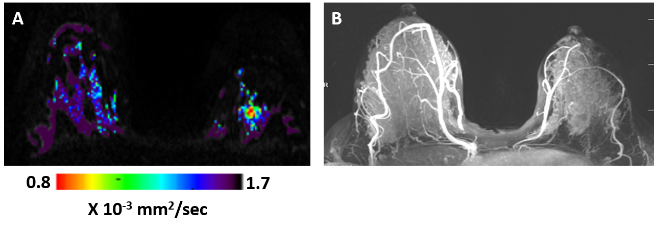
Figure 2: DTI λ1 parametric map (A) and maximum intensity projection (MIP) of the DCE images (B) in a lactating 33 years old BRCA-1 carrier woman. The λ1 parametric map depicted in the left breast a non-palpable invasive ductal carcinoma lesion (low λ1 values) that was detected by screening DTI but was not demonstrated by DCE because of the masking effect of marked background parenchymal enhancement (BIRADS 1 DCE and BIRADS 5 DTI).
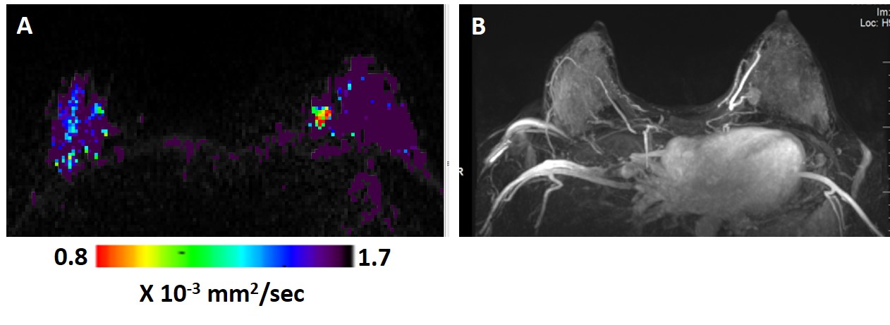
Figure 1: DTI λ1 parametric map (A) and maximum intensity projection (MIP) of the DCE images (B) in a lactating 33 years old breast cancer patient. The λ1 parametric map and the DCE images both show in the left breast a triple negative invasive ductal carcinoma mass lesion (low λ1 values ). The cancer lesion was diagnosed by both DCE (assigned BIRADS 4) and by DTI (assigned BIRADS 5).