Ping N Wang1, Julia V Velikina2, Alexey A Samsonov2, Lloyd Estkowski3, Ty A Cashen3, Frederick Felcz2, Roberta M Strigel1,2,4, Frank R Korosec1,2, and James H Holmes2
1Department of Medical Physics, University of Wisconsin-Madison, Madison, WI, United States, 2Department of Radiology, University of Wisconsin-Madison, Madison, WI, United States, 3Global MR Applications & Workflow, GE Healthcare, Madison, WI, United States, 4Carbone Cancer Center, University of Wisconsin-Madison, Madison, WI, United States
1Department of Medical Physics, University of Wisconsin-Madison, Madison, WI, United States, 2Department of Radiology, University of Wisconsin-Madison, Madison, WI, United States, 3Global MR Applications & Workflow, GE Healthcare, Madison, WI, United States, 4Carbone Cancer Center, University of Wisconsin-Madison, Madison, WI, United States
This work demonstrates the feasibility of providing
motion-free high spatial-temporal resolution breast DCE-MRI using radial
acquisition combined with self-gating and MOCCO reconstruction.
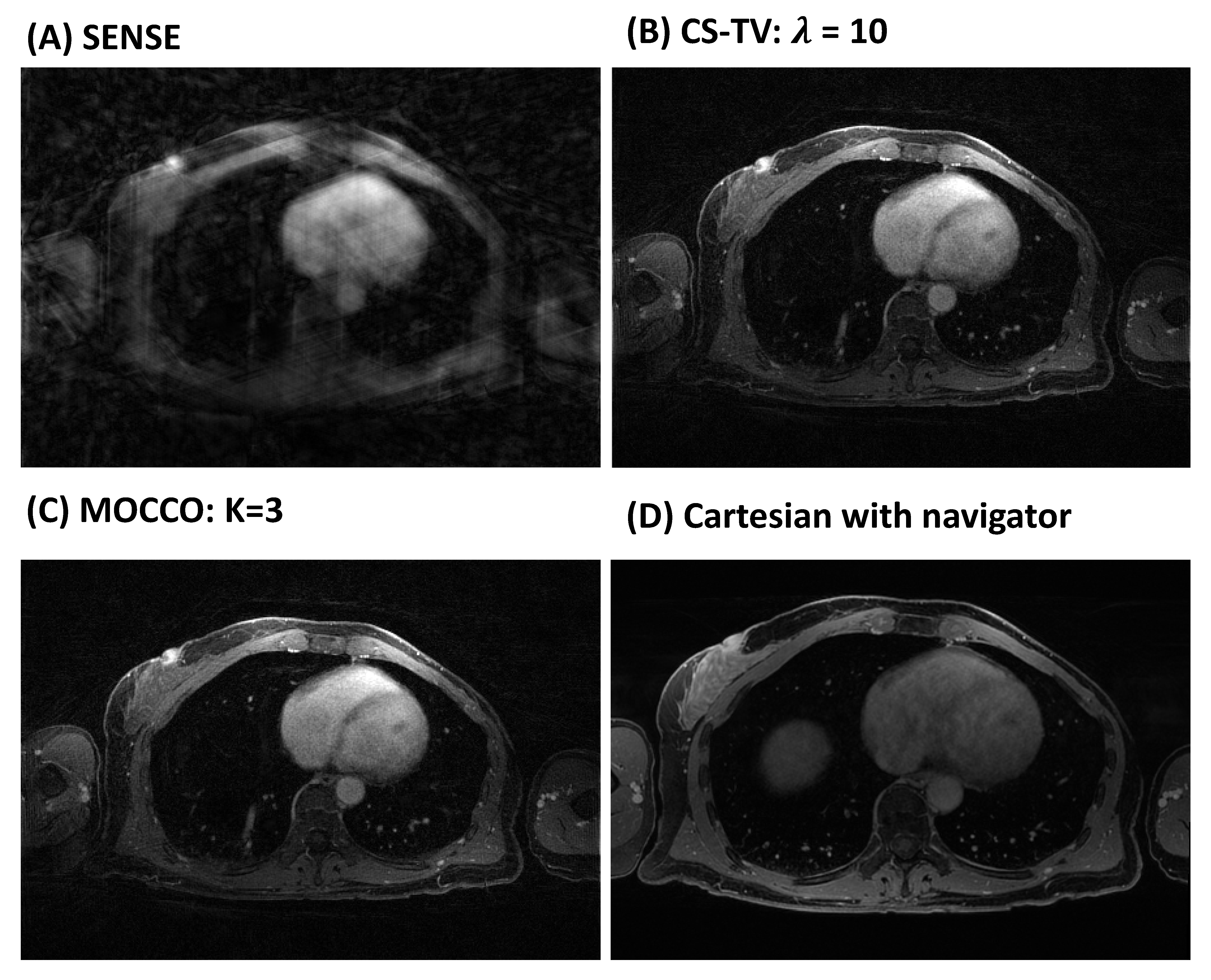
Figure
4: Individual time frames at peak contrast enhancement (time = 240 s)
reconstructed using SENSE (A), CS-TV (B), MOCCO with model order K=3 (C) with
temporal resolution of 15 s. High image quality is observed for both
regularized reconstructions (B, C). Note the late-phase image (D) was acquired
with a navigator-gated Cartesian acquisition, which resulted in a longer acquisition
time (8:43 min:s).
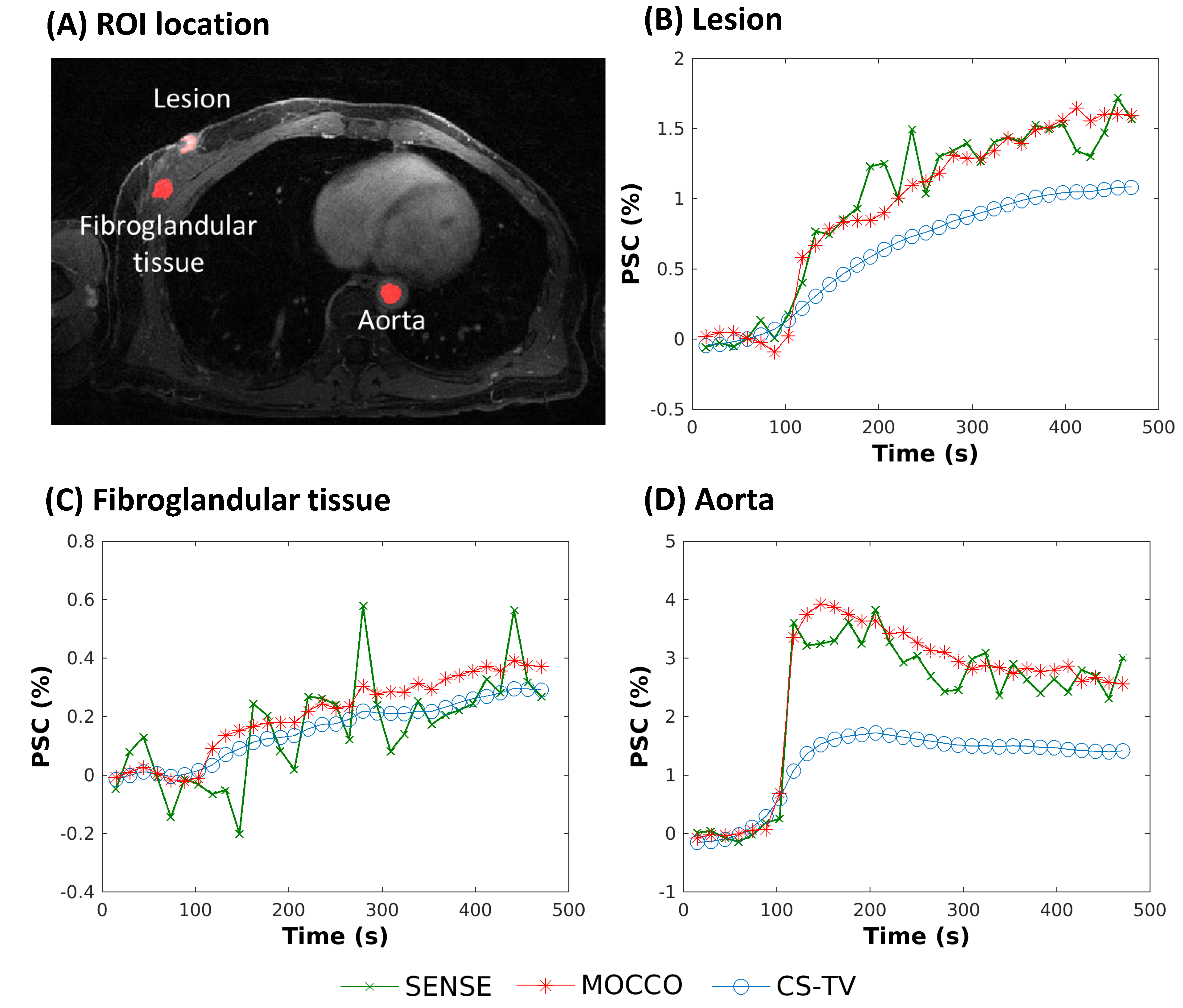
Figure
5: Supine image (A) from a patient volunteer reconstructed using SENSE (green,
X), CS-TV (blue, circles), and MOCCO with model order K=3 (red, stars) with 15
s temporal resolution. PSC (%) are plotted from ROIs placed in the lesion,
fibroglandular tissue, and aorta (A, red outlines). Rapid wash-in and wash-out
contrast kinetics are observed in the aorta D). The lesion B) showed relatively
rapid contrast update, while the fibroglandular tissue C) showed slower
contrast uptake.