1Institute of Medical Sciences, University of Aberdeen, Aberdeen, United Kingdom, 2Pathology Department, Aberdeen Royal Infirmary, Aberdeen, United Kingdom, 3School of Medicine, University of Aberdeen, Aberdeen, United Kingdom, 4Breast Unit, Aberdeen Royal Infirmary, Aberdeen, United Kingdom
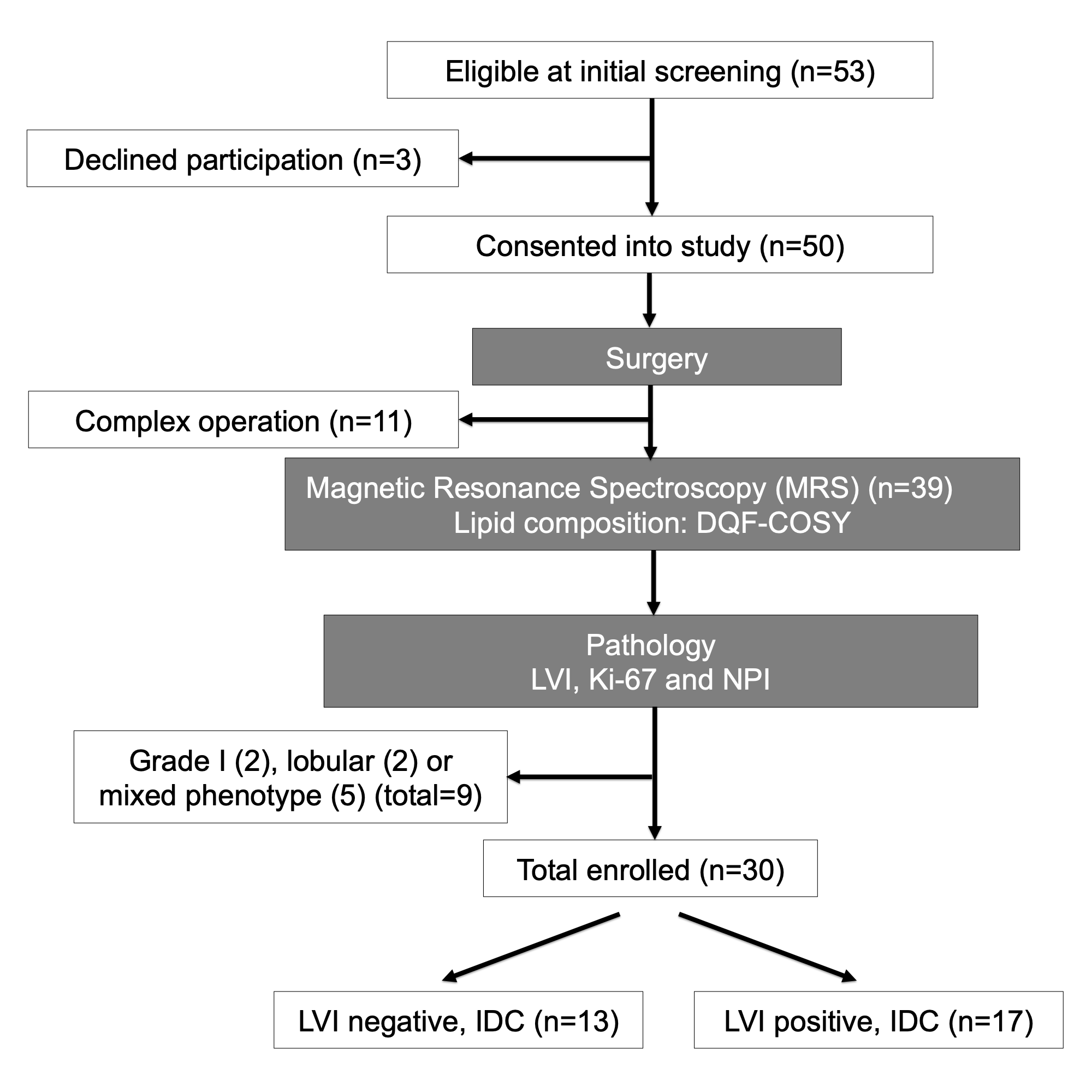
Figure 1. Study design.
A two-group cross sectional study in a flow chart. Freshly excised breast tumours from wide local excision or mastectomy were immediately scanned on a clinical 3 T MRI scanner to derive lipid composition using double quantum filtered–correlation spectroscopy (DQF-COSY). Immunohistochemical examinations were conducted to assess lymphovascular invasion (LVI), Ki-67 expression and Nottingham Prognostic Index (NPI). In total, 30 patients with invasive ductal carcinoma (IDC), 13 with LVI negative and 17 with LVI positive, participated in the study.
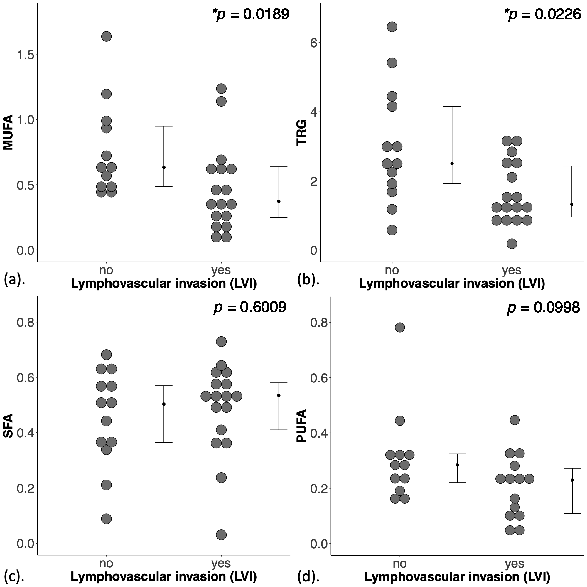
Figure 2. Group difference in lipid composition.
The difference in (a) monounsaturated fatty acids (MUFA), (b) triglycerides (TRG), (c) saturated FA (SFA) and (d) polyunsaturated FA (PUFA), are shown in dot plots. Each dot represents the measurement from a patient, and the dots are organised in two columns corresponding to the lymphovascular invasion (LVI) status. The error bar indicates the median and interquartile range. The Mann Whitney U tests were performed between the groups and p value is shown for each plot. Statistically significant p values (< 0.05) are marked by ‘*’.