Ioannis Papadopoulos1, Ivan Dimitrov2, Jochen Keupp3, Durga Udayakumar1,4, Stephen Seiler1, Sunati Sahoo5, Yin Xi1, Emily Knippa1, Robert Lenkinski1,4, Ananth Madhuranthakam1,4, Shu Zhang6, and Elena Vinogradov1,4
1Radiology, University of Texas Southwestern Medical Center, Dallas, TX, United States, 2Philips Healthcare, Gainesville, FL, United States, 3Philips Research, Hamburg, Germany, 4Advanced Imaging Research Center, University of Texas Southwestern Medical Center, Dallas, TX, United States, 5Pathology, University of Texas Southwestern Medical Center, Dallas, TX, United States, 6Cancer Systems Imaging, University of Texas MD Anderson Cancer Center, Houston, TX, United States
1Radiology, University of Texas Southwestern Medical Center, Dallas, TX, United States, 2Philips Healthcare, Gainesville, FL, United States, 3Philips Research, Hamburg, Germany, 4Advanced Imaging Research Center, University of Texas Southwestern Medical Center, Dallas, TX, United States, 5Pathology, University of Texas Southwestern Medical Center, Dallas, TX, United States, 6Cancer Systems Imaging, University of Texas MD Anderson Cancer Center, Houston, TX, United States
CEST-mDixon using MTRasym at 1ppm and 2ppm, shows significant potential in breast tumor aggressiveness
differentiation. Ki-67 correlation is confirmed and, for the first time, a
negative linear correlation with the percentage of cells positive for nuclear
expression of the PR is found.
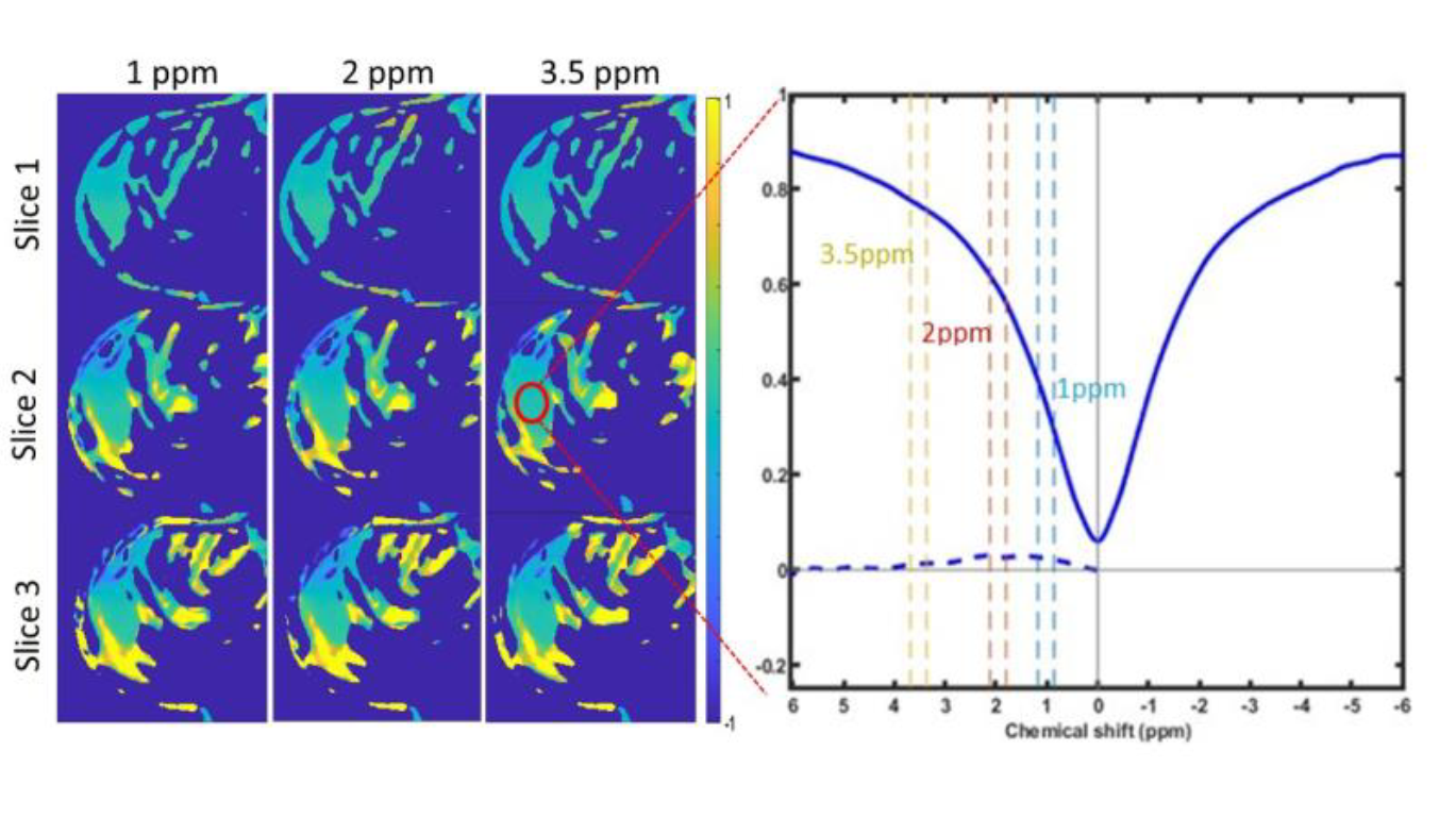
CEST-mDixon result. Left: MTRasym maps for 1ppm, 2ppm and
3.5ppm for 3 slices. Right: Z-spectrum from the ROI placed over the suspicious
lesion.
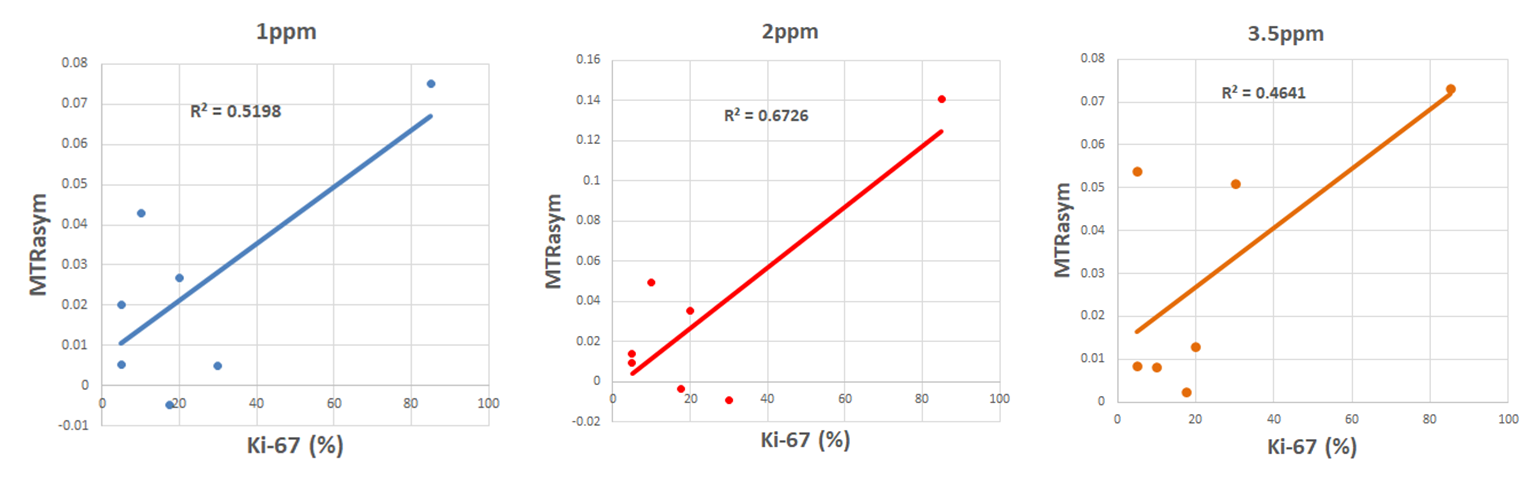
Left to right: ROI averaged MTRasym(1ppm), MTRasym(2ppm)
and MTRasym(3.5ppm) compared to the Ki-67 histopathological index.
A moderate positive correlation is observed for hydroxyls (1ppm) and amines (2
ppm).