Olivia C Sehl1,2 and Paula J Foster1,2
1Imaging Research Laboratories, Robarts Research Institute, London, ON, Canada, 2Medical Biophysics, University of Western Ontario, London, ON, Canada
1Imaging Research Laboratories, Robarts Research Institute, London, ON, Canada, 2Medical Biophysics, University of Western Ontario, London, ON, Canada
The detection of stem cells using 19F MRI and MPI with common cell labeling agents (perfluorocarbon and ferucarbotran) was evaluated. There were significant increases in 19F and MPI signal with cell number and fewer cells were detected with MPI.

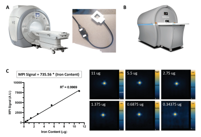
Figure 1: A picture of the (A) 3 Tesla clinical MRI (MR750, General Electric) and dual-tuned (1H/19F) surface coil (MR Solutions) used for 19F imaging, and (B) preclinical MomentumTM MPI scanner (Magnetic Insight Inc.). (C) The relationship between MPI signal and iron mass is determined for MPI signal calibration by imaging known amounts of ferucarbotran with 3.0 T/m gradients.

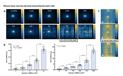
Figure 4: MPI detection of ferucarbotran-labeled MSC. (A) 2D MPI of individual MSC pellets containing various cell numbers (M = 106, k = 103). As few as 2000 MSC was detected in 6 of 9 replicates, however with low SNR (2.51). The detection limit of 4000 MSC is reliable and quantifiable (9/9 replicates, SNR = 3.77). (B) 2D MPI signal (and the associated iron content) significantly increases with number of MSC. (D) In 3D (35 projections), 2000 cells can be reliably detected (9/9 replicates, SNR = 6.42) and as few as 1000 cells were detected in 2 of 9 replicates (SNR = 4.69, signal volume 116mm3).
