Kianush Karimian-Jazi1, Ulf Neuberger1, Katharina Schregel1, Gianluca Brugnara1, Daniel Schwarz1, Laura Jäger2, Wolfgang Wick2, Martin Bendszus1, and Michael Oliver Breckwoldt1
1Neuroradiology, University Clinic of Heidelberg, Heidelberg, Germany, 2Neurology, University Clinic of Heidelberg, Heidelberg, Germany
1Neuroradiology, University Clinic of Heidelberg, Heidelberg, Germany, 2Neurology, University Clinic of Heidelberg, Heidelberg, Germany
Gd-contrast administration is dispensable in follow-up spinal MRI of MS patients if no new T2 lesions are present. We show high sensitivity and specificity of the T2 signal ratio as a predictor for enhancing, “active” lesions and propose a shortened, single MRI protocol for routine follow-up MRI
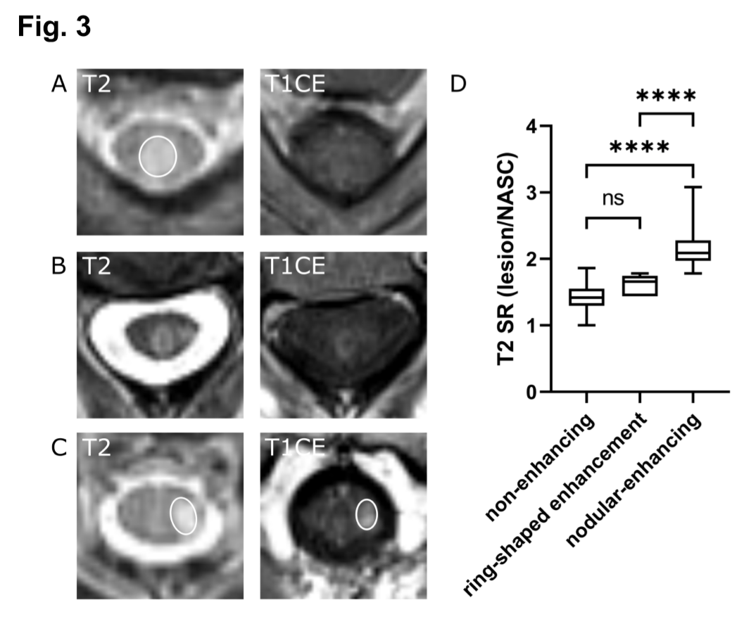
Fig. 3 Representative MRI images of axial T2-w and T1-w CE images showing different lesion morphologies. (A) T2 lesion without corresponding enhancement and a low T2 signal ratio (SR: 1.54). (B) Ring-enhancing lesion with a T2 SR of 1.78. (C) T2 lesion with a high T2 signal ratio (SR: 1.93) and clear contrast-enhancement. (D) Nodularenhancing lesions (n=33) showed significantly higher T2 signal ratios than nonenhancing (n=185, ****p<0.0001) and ring-shaped enhancing lesions (n=5 ****p<0.0001). NASC: normal appearing spinal cord.
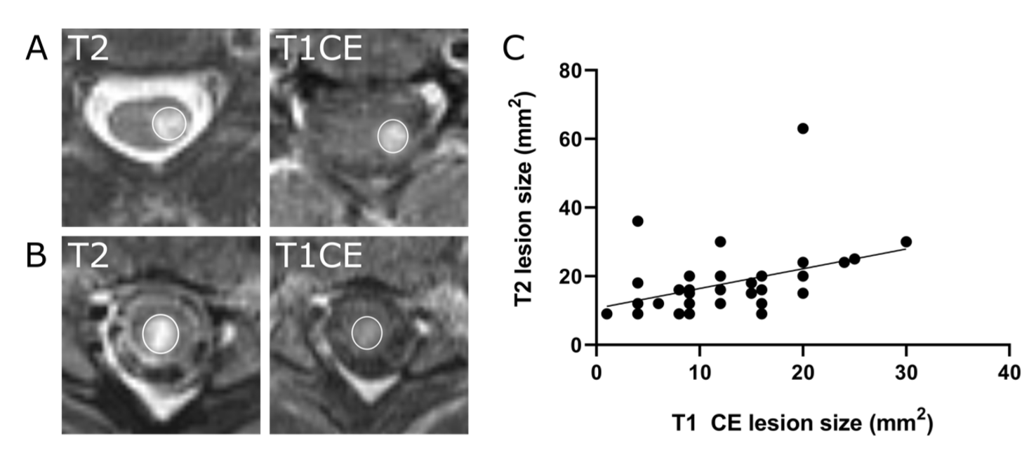
Fig. 2 T2-w and T1-w contrast enhanced images (A, B). Size of the T2 lesion weakly correlates with the size of the contrast enhancement (C). Pearson correlation analysis, r2 = 0.17, **p < 0.01, n = 44 lesions).